Show PDB file:   
         Plain Text   HTML   (compressed file size)
QuickSearch:   
by PDB,NDB,UniProt,PROSITE Code or Search Term(s)  
(-)Asymmetric Unit
(-)Asym. Unit - sites
(-)Biological Unit 1
(-)Biol. Unit 1 - sites
(-)Biological Unit 2
collapse expand < >
Image Asymmetric Unit
Asymmetric Unit  (Jmol Viewer)
Image Asym. Unit - sites
Asym. Unit - sites  (Jmol Viewer)
Image Biological Unit 1
Biological Unit 1  (Jmol Viewer)
Image Biol. Unit 1 - sites
Biol. Unit 1 - sites  (Jmol Viewer)
Image Biological Unit 2
Biological Unit 2  (Jmol Viewer)

(-) Description

Title :  CEL6A IN COMPLEX WITH M-IODOBENZYL BETA-D-GLUCOPYRANOSYL-BETA(1,4)-D-XYLOPYRANOSIDE
 
Authors :  J. -Y. Zou, T. A. Jones
Date :  08 Jul 99  (Deposition) - 18 Sep 99  (Release) - 27 Jan 16  (Revision)
Method :  X-RAY DIFFRACTION
Resolution :  2.10
Chains :  Asym. Unit :  A,B
Biol. Unit 1:  A  (1x)
Biol. Unit 2:  B  (1x)
Keywords :  Hydrolase, Glycosidase, Glycoprotein (Keyword Search: [Gene Ontology, PubMed, Web (Google))
 
Reference :  J. -Y. Zou, G. J. Kleywegt, J. Stahlberg, H. Driguez, W. Nerinckx, M. Claeyssens, A. Koivula, T. T. Teeri, T. A. Jones
Crystallographic Evidence For Substrate Ring Distortion And Protein Conformational Changes During Catalysis In Cellobiohydrolase Cel6A From Trichoderma Reesei
Structure V. 7 1035 1999
PubMed-ID: 10508787  |  Reference-DOI: 10.1016/S0969-2126(99)80171-3
(for further references see the PDB file header)

(-) Compounds

Molecule 1 - CELLOBIOHYDROLASE CEL6A (FORMERLY CALLED CBH II)
    ChainsA, B
    EC Number3.2.1.91
    EngineeredYES
    Expression SystemTRICHODERMA REESEI
    Expression System Taxid51453
    FragmentCATALYTIC DOMAIN, RESIDUES 85-447
    Organism ScientificTRICHODERMA REESEI
    Organism Taxid51453
    SynonymCEL6A

 Structural Features

(-) Chains, Units

  12
Asymmetric Unit AB
Biological Unit 1 (1x)A 
Biological Unit 2 (1x) B

Summary Information (see also Sequences/Alignments below)

(-) Ligands, Modified Residues, Ions  (9, 27)

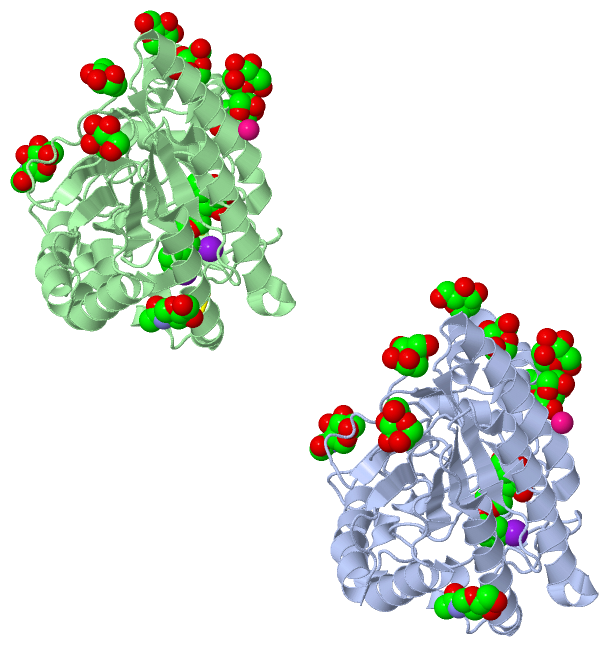
Asymmetric Unit (9, 27)
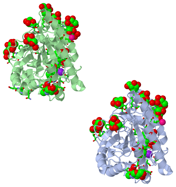
No.NameCountTypeFull Name
1BGC1Ligand/IonBETA-D-GLUCOSE
2CO2Ligand/IonCOBALT (II) ION
3GLC1Ligand/IonALPHA-D-GLUCOSE
4IOB2Ligand/Ion3-IODO-BENZYL ALCOHOL
5IOD1Ligand/IonIODIDE ION
6MAN14Ligand/IonALPHA-D-MANNOSE
7NAG4Ligand/IonN-ACETYL-D-GLUCOSAMINE
8XYP1Ligand/IonBETA-D-XYLOPYRANOSE
9XYS1Ligand/IonXYLOPYRANOSE
Biological Unit 1 (5, 12)
No.NameCountTypeFull Name
1BGC1Ligand/IonBETA-D-GLUCOSE
2CO-1Ligand/IonCOBALT (II) ION
3GLC-1Ligand/IonALPHA-D-GLUCOSE
4IOB1Ligand/Ion3-IODO-BENZYL ALCOHOL
5IOD-1Ligand/IonIODIDE ION
6MAN7Ligand/IonALPHA-D-MANNOSE
7NAG2Ligand/IonN-ACETYL-D-GLUCOSAMINE
8XYP1Ligand/IonBETA-D-XYLOPYRANOSE
9XYS-1Ligand/IonXYLOPYRANOSE
Biological Unit 2 (6, 13)
No.NameCountTypeFull Name
1BGC-1Ligand/IonBETA-D-GLUCOSE
2CO-1Ligand/IonCOBALT (II) ION
3GLC1Ligand/IonALPHA-D-GLUCOSE
4IOB1Ligand/Ion3-IODO-BENZYL ALCOHOL
5IOD1Ligand/IonIODIDE ION
6MAN7Ligand/IonALPHA-D-MANNOSE
7NAG2Ligand/IonN-ACETYL-D-GLUCOSAMINE
8XYP-1Ligand/IonBETA-D-XYLOPYRANOSE
9XYS1Ligand/IonXYLOPYRANOSE

(-) Sites  (23, 23)

Asymmetric Unit (23, 23)
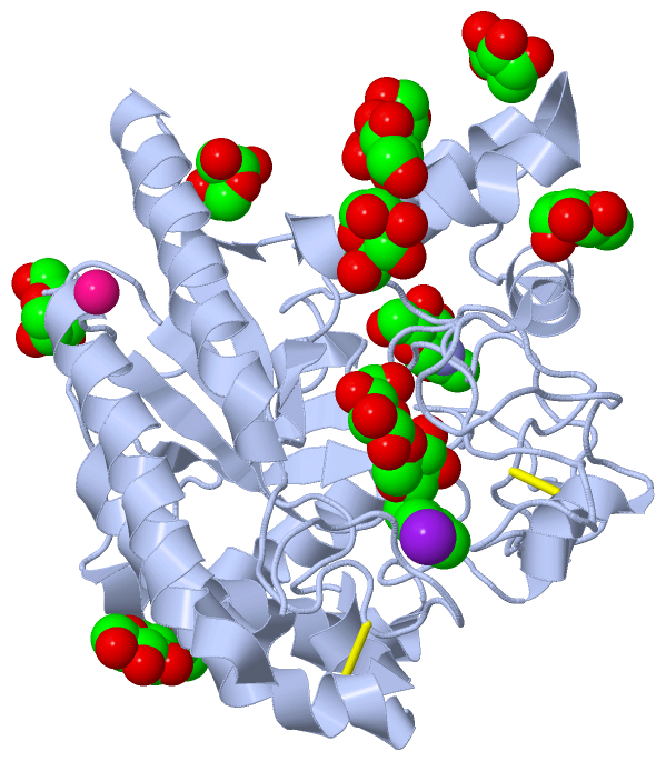
No.NameEvidenceResiduesDescription
01AC1SOFTWAREGLU A:146BINDING SITE FOR RESIDUE CO A 900
02AC2SOFTWAREALA B:178 , ALA B:180 , IOB B:901BINDING SITE FOR RESIDUE IOD B 899
03AC3SOFTWAREGLU B:146BINDING SITE FOR RESIDUE CO B 900
04AC4SOFTWARETHR A:87 , LYS A:356BINDING SITE FOR MONO-SACCHARIDE MAN A 503 BOUND TO THR A 87
05AC5SOFTWARETHR A:97 , ASN A:161BINDING SITE FOR MONO-SACCHARIDE MAN A 504 BOUND TO THR A 97
06AC6SOFTWARETYR A:103 , SER A:106 , LEU A:111 , MAN A:507BINDING SITE FOR MONO-SACCHARIDE MAN A 505 BOUND TO SER A 106
07AC7SOFTWAREALA A:105 , SER A:109 , ALA A:125 , ALA A:128 , LYS A:129 , HOH A:2264 , HOH A:2265 , ASN B:339BINDING SITE FOR MONO-SACCHARIDE MAN A 506 BOUND TO SER A 109
08AC8SOFTWARESER A:106 , SER A:110 , MAN A:505 , HOH A:2266 , GLN B:281 , HIS B:340BINDING SITE FOR MONO-SACCHARIDE MAN A 507 BOUND TO SER A 110
09AC9SOFTWARESER A:115 , GLN A:432 , HOH A:2250 , HOH A:2268 , HOH A:2269 , HOH A:2270 , HOH A:2271BINDING SITE FOR MONO-SACCHARIDE MAN A 508 BOUND TO SER A 115
10BC1SOFTWAREGLY A:118 , THR A:122BINDING SITE FOR MONO-SACCHARIDE MAN A 509 BOUND TO THR A 122
11BC2SOFTWARELEU A:243 , GLU A:244 , ASN A:247 , ASN A:289 , HOH A:2260 , HOH A:2261BINDING SITE FOR MONO-SACCHARIDE NAG A 501 BOUND TO ASN A 289
12BC3SOFTWARETRP A:309 , ASN A:310 , THR A:312 , PHE A:391 , HOH A:2163 , HOH A:2263BINDING SITE FOR MONO-SACCHARIDE NAG A 502 BOUND TO ASN A 310
13BC4SOFTWARETHR B:87 , GLY B:372 , ASN B:441 , HOH B:2227BINDING SITE FOR MONO-SACCHARIDE MAN B 503 BOUND TO THR B 87
14BC5SOFTWARETHR B:97 , ASN B:161BINDING SITE FOR MONO-SACCHARIDE MAN B 504 BOUND TO THR B 97
15BC6SOFTWARETYR B:103 , SER B:106 , LEU B:111 , MAN B:507 , HOH B:2228 , HOH B:2229BINDING SITE FOR MONO-SACCHARIDE MAN B 505 BOUND TO SER B 106
16BC7SOFTWAREALA B:105 , SER B:109 , ALA B:125 , LYS B:129 , HOH B:2230BINDING SITE FOR MONO-SACCHARIDE MAN B 506 BOUND TO SER B 109
17BC8SOFTWARESER B:106 , SER B:110 , MAN B:505BINDING SITE FOR MONO-SACCHARIDE MAN B 507 BOUND TO SER B 110
18BC9SOFTWARESER B:115 , GLN B:432 , HOH B:2232BINDING SITE FOR MONO-SACCHARIDE MAN B 508 BOUND TO SER B 115
19CC1SOFTWAREGLY B:118 , THR B:122BINDING SITE FOR MONO-SACCHARIDE MAN B 509 BOUND TO THR B 122
20CC2SOFTWARELEU B:243 , GLU B:244 , ASN B:247 , ASN B:289 , HOH B:2224BINDING SITE FOR MONO-SACCHARIDE NAG B 501 BOUND TO ASN B 289
21CC3SOFTWARETRP B:309 , ASN B:310 , THR B:312 , PHE B:391 , HOH B:2181 , HOH B:2225 , HOH B:2226BINDING SITE FOR MONO-SACCHARIDE NAG B 502 BOUND TO ASN B 310
22CC4SOFTWARETRP A:135 , ASP A:137 , TYR A:169 , ALA A:178 , ALA A:180 , ASP A:221 , TRP A:367 , LYS A:395 , GLU A:399 , ASP A:401 , HOH A:2066 , HOH A:2117 , HOH A:2159 , HOH A:2221 , HOH A:2273 , HOH A:2274 , HOH A:2275BINDING SITE FOR POLY-SACCHARIDE RESIDUES IOB A 901 THROUGH BGC A 903
23CC5SOFTWARETRP A:135 , ASP A:137 , TYR A:169 , ALA A:178 , ALA A:180 , ASP A:221 , TRP A:367 , LYS A:395 , GLU A:399 , ASP A:401 , HOH A:2066 , HOH A:2117 , HOH A:2159 , HOH A:2221 , HOH A:2273 , HOH A:2274 , HOH A:2275 , TRP B:135 , ASP B:137 , TYR B:169 , ASP B:221 , TRP B:367 , LYS B:395 , GLU B:399 , ASP B:401 , IOD B:899 , HOH B:2131 , HOH B:2233 , HOH B:2234 , HOH B:2235 , HOH B:2236BINDING SITE FOR POLY-SACCHARIDE RESIDUES IOB B 901 THROUGH GLC B 903

(-) SS Bonds  (4, 4)

Asymmetric Unit
No.Residues
1A:176 -A:235
2A:368 -A:415
3B:176 -B:235
4B:368 -B:415

(-) Cis Peptide Bonds  (6, 6)

Asymmetric Unit
No.Residues
1Gln A:357 -Pro A:358
2Gln A:422 -Pro A:423
3Asn A:443 -Pro A:444
4Gln B:357 -Pro B:358
5Gln B:422 -Pro B:423
6Asn B:443 -Pro B:444

 Sequence-Structure Mapping

(-) SAPs(SNPs)/Variants  (0, 0)

(no "SAP(SNP)/Variant" information available for 1QK0)

(-) PROSITE Motifs  (2, 4)

Asymmetric Unit (2, 4)
 PROSITEUniProtKBPDB
No.IDACDescriptionIDLocationCountLocation
1GLYCOSYL_HYDROL_F6_1PS00655 Glycosyl hydrolases family 6 signature 1.GUX2_HYPJE191-207
 
  2A:167-183
B:167-183
2GLYCOSYL_HYDROL_F6_2PS00656 Glycosyl hydrolases family 6 signature 2.GUX2_HYPJE239-248
 
  2A:215-224
B:215-224
Biological Unit 1 (2, 2)
 PROSITEUniProtKBPDB
No.IDACDescriptionIDLocationCountLocation
1GLYCOSYL_HYDROL_F6_1PS00655 Glycosyl hydrolases family 6 signature 1.GUX2_HYPJE191-207
 
  1A:167-183
-
2GLYCOSYL_HYDROL_F6_2PS00656 Glycosyl hydrolases family 6 signature 2.GUX2_HYPJE239-248
 
  1A:215-224
-
Biological Unit 2 (2, 2)
 PROSITEUniProtKBPDB
No.IDACDescriptionIDLocationCountLocation
1GLYCOSYL_HYDROL_F6_1PS00655 Glycosyl hydrolases family 6 signature 1.GUX2_HYPJE191-207
 
  1-
B:167-183
2GLYCOSYL_HYDROL_F6_2PS00656 Glycosyl hydrolases family 6 signature 2.GUX2_HYPJE239-248
 
  1-
B:215-224

(-) Exons   (0, 0)

(no "Exon" information available for 1QK0)

(-) Sequences/Alignments

Asymmetric Unit
   Reformat: Number of residues per line =  ('0' or empty: single-line sequence representation)
  Number of residues per labelling interval =   
  UniProt sequence: complete  aligned part    
   Show mapping: SCOP domains CATH domains Pfam domains Secondary structure (by author)
SAPs(SNPs) PROSITE motifs Exons
(details for a mapped element are shown in a popup box when the mouse pointer rests over it)
Chain A from PDB  Type:PROTEIN  Length:363
 aligned with GUX2_HYPJE | P07987 from UniProtKB/Swiss-Prot  Length:471

    Alignment length:363
                                   118       128       138       148       158       168       178       188       198       208       218       228       238       248       258       268       278       288       298       308       318       328       338       348       358       368       378       388       398       408       418       428       438       448       458       468   
           GUX2_HYPJE   109 TATYSGNPFVGVTPWANAYYASEVSSLAIPSLTGAMATAAAAVAKVPSFMWLDTLDKTPLMEQTLADIRTANKNGGNYAGQFVVYDLPDRDCAALASNGEYSIADGGVAKYKNYIDTIRQIVVEYSDIRTLLVIEPDSLANLVTNLGTPKCANAQSAYLECINYAVTQLNLPNVAMYLDAGHAGWLGWPANQDPAAQLFANVYKNASSPRALRGLATNVANYNGWNITSPPSYTQGNAVYNEKLYIHAIGPLLANHGWSNAFFITDQGRSGKQPTGQQQWGDWCNVIGTGFGIRPSANTGDSLLDSFVWVKPGGECDGTSDSSAPRFDSHCALPDALQPAPQAGAWFQAYFVQLLTNANPSFL 471
               SCOP domains d1qk0a_ A: Cellobiohydrolase II (Cel6)                                                                                                                                                                                                                                                                                                                                      SCOP domains
               CATH domains 1qk0A00 A:85-447 7-stranded glycosidases (cellulases)                                                                                                                                                                                                                                                                                                                       CATH domains
               Pfam domains --------------------------------------------------------------------------------------------------------------------------------------------------------------------------------------------------------------------------------------------------------------------------------------------------------------------------------------------------------------------------- Pfam domains
         Sec.struct. author ............ee..hhhhhhhhhhhhhhhhhhhhhhhhhhhh.....eee.hhhhhhhhhhhhhhhhhhhhh...eeeeeee.................hhhhhhhhhhhhhhhhhhhhhhhh....eeeee.....hhhhhh..hhhhhhhhhhhhhhhhhhhhhh....eeeeee........hhhhhhhhhhhhhhhhhhh.....eeeeee.............hhhhh.....hhhhhhhhhhhhhhhh.....eeeee.....................................eeeee...................hhhhhh.............hhhhhhhhhhh...... Sec.struct. author
                 SAPs(SNPs) --------------------------------------------------------------------------------------------------------------------------------------------------------------------------------------------------------------------------------------------------------------------------------------------------------------------------------------------------------------------------- SAPs(SNPs)
                    PROSITE ----------------------------------------------------------------------------------GLYCOSYL_HYDROL_F-------------------------------GLYCOSYL_H------------------------------------------------------------------------------------------------------------------------------------------------------------------------------------------------------------------------------- PROSITE
                 Transcript --------------------------------------------------------------------------------------------------------------------------------------------------------------------------------------------------------------------------------------------------------------------------------------------------------------------------------------------------------------------------- Transcript
                 1qk0 A  85 TATYSGNPFVGVTPWANAYYASEVSSLAIPSLTGAMATAAAAVAKVPSFMWLDTLDKTPLMEQTLADIRTANKNGGNYAGQFVVYDLPDRDCAALASNGEYSIADGGVAKYKNYIDTIRQIVVEYSDIRTLLVIEPDSLANLVTNLGTPKCANAQSAYLECINYAVTQLNLPNVAMYLDAGHAGWLGWPANQDPAAQLFANVYKNASSPRALRGLATNVANYNGWNITSPPSYTQGNAVYNEKLYIHAIGPLLANHGWSNAFFITDQGRSGKQPTGQQQWGDWCNVIGTGFGIRPSANTGDSLLDSFVWVKPGGECDGTSDSSAPRFDSHCALPDALQPAPQAGAWFQAYFVQLLTNANPSFL 447
                                    94       104       114       124       134       144       154       164       174       184       194       204       214       224       234       244       254       264       274       284       294       304       314       324       334       344       354       364       374       384       394       404       414       424       434       444   

Chain B from PDB  Type:PROTEIN  Length:363
 aligned with GUX2_HYPJE | P07987 from UniProtKB/Swiss-Prot  Length:471

    Alignment length:363
                                   118       128       138       148       158       168       178       188       198       208       218       228       238       248       258       268       278       288       298       308       318       328       338       348       358       368       378       388       398       408       418       428       438       448       458       468   
           GUX2_HYPJE   109 TATYSGNPFVGVTPWANAYYASEVSSLAIPSLTGAMATAAAAVAKVPSFMWLDTLDKTPLMEQTLADIRTANKNGGNYAGQFVVYDLPDRDCAALASNGEYSIADGGVAKYKNYIDTIRQIVVEYSDIRTLLVIEPDSLANLVTNLGTPKCANAQSAYLECINYAVTQLNLPNVAMYLDAGHAGWLGWPANQDPAAQLFANVYKNASSPRALRGLATNVANYNGWNITSPPSYTQGNAVYNEKLYIHAIGPLLANHGWSNAFFITDQGRSGKQPTGQQQWGDWCNVIGTGFGIRPSANTGDSLLDSFVWVKPGGECDGTSDSSAPRFDSHCALPDALQPAPQAGAWFQAYFVQLLTNANPSFL 471
               SCOP domains d1qk0b_ B: Cellobiohydrolase II (Cel6)                                                                                                                                                                                                                                                                                                                                      SCOP domains
               CATH domains 1qk0B00 B:85-447 7-stranded glycosidases (cellulases)                                                                                                                                                                                                                                                                                                                       CATH domains
               Pfam domains --------------------------------------------------------------------------------------------------------------------------------------------------------------------------------------------------------------------------------------------------------------------------------------------------------------------------------------------------------------------------- Pfam domains
         Sec.struct. author ............ee..hhhhhhhhhhhhhhhhhhhhhhhhhhhh.....eee.hhhhhhhhhhhhhhhhhhhhh...eeeeeee.................hhhhhhhhhhhhhhhhhhhhhhhh....eeeee...hhhhhhhh..hhhhhhhhhhhhhhhhhhhhhh....eeeeee..hhhhhhhhhhhhhhhhhhhhhhhhh.....eeeeee.............hhhhh.....hhhhhhhhhhhhhhhh.....eeeee.....................................eeeee...................hhhhhh.............hhhhhhhhhhh...... Sec.struct. author
                 SAPs(SNPs) --------------------------------------------------------------------------------------------------------------------------------------------------------------------------------------------------------------------------------------------------------------------------------------------------------------------------------------------------------------------------- SAPs(SNPs)
                    PROSITE ----------------------------------------------------------------------------------GLYCOSYL_HYDROL_F-------------------------------GLYCOSYL_H------------------------------------------------------------------------------------------------------------------------------------------------------------------------------------------------------------------------------- PROSITE
                 Transcript --------------------------------------------------------------------------------------------------------------------------------------------------------------------------------------------------------------------------------------------------------------------------------------------------------------------------------------------------------------------------- Transcript
                 1qk0 B  85 TATYSGNPFVGVTPWANAYYASEVSSLAIPSLTGAMATAAAAVAKVPSFMWLDTLDKTPLMEQTLADIRTANKNGGNYAGQFVVYDLPDRDCAALASNGEYSIADGGVAKYKNYIDTIRQIVVEYSDIRTLLVIEPDSLANLVTNLGTPKCANAQSAYLECINYAVTQLNLPNVAMYLDAGHAGWLGWPANQDPAAQLFANVYKNASSPRALRGLATNVANYNGWNITSPPSYTQGNAVYNEKLYIHAIGPLLANHGWSNAFFITDQGRSGKQPTGQQQWGDWCNVIGTGFGIRPSANTGDSLLDSFVWVKPGGECDGTSDSSAPRFDSHCALPDALQPAPQAGAWFQAYFVQLLTNANPSFL 447
                                    94       104       114       124       134       144       154       164       174       184       194       204       214       224       234       244       254       264       274       284       294       304       314       324       334       344       354       364       374       384       394       404       414       424       434       444   

   Legend:   → Mismatch (orange background)
  - → Gap (green background, '-', border residues have a numbering label)
    → Modified Residue (blue background, lower-case, 'x' indicates undefined single-letter code, labelled with number + name)
  x → Chemical Group (purple background, 'x', labelled with number + name, e.g. ACE or NH2)
  extra numbering lines below/above indicate numbering irregularities and modified residue names etc., number ends below/above '|'

 Classification and Annotation

(-) SCOP Domains  (1, 2)

Asymmetric Unit

(-) CATH Domains  (1, 2)

Asymmetric Unit
(-)
Class: Alpha Beta (26913)

(-) Pfam Domains  (0, 0)

(no "Pfam Domain" information available for 1QK0)

(-) Gene Ontology  (11, 11)

Asymmetric Unit(hide GO term definitions)
Chain A,B   (GUX2_HYPJE | P07987)
molecular function
    GO:0016162    cellulose 1,4-beta-cellobiosidase activity    Catalysis of the hydrolysis of (1->4)-beta-D-glucosidic linkages in cellulose and cellotetraose, releasing cellobiose from the non-reducing ends of the chains.
    GO:0030248    cellulose binding    Interacting selectively and non-covalently with cellulose.
    GO:0016787    hydrolase activity    Catalysis of the hydrolysis of various bonds, e.g. C-O, C-N, C-C, phosphoric anhydride bonds, etc. Hydrolase is the systematic name for any enzyme of EC class 3.
    GO:0016798    hydrolase activity, acting on glycosyl bonds    Catalysis of the hydrolysis of any glycosyl bond.
    GO:0004553    hydrolase activity, hydrolyzing O-glycosyl compounds    Catalysis of the hydrolysis of any O-glycosyl bond.
    GO:0042802    identical protein binding    Interacting selectively and non-covalently with an identical protein or proteins.
biological process
    GO:0005975    carbohydrate metabolic process    The chemical reactions and pathways involving carbohydrates, any of a group of organic compounds based of the general formula Cx(H2O)y. Includes the formation of carbohydrate derivatives by the addition of a carbohydrate residue to another molecule.
    GO:0030245    cellulose catabolic process    The chemical reactions and pathways resulting in the breakdown of cellulose, a linear beta1-4 glucan of molecular mass 50-400 kDa with the pyranose units in the -4C1 conformation.
    GO:0008152    metabolic process    The chemical reactions and pathways, including anabolism and catabolism, by which living organisms transform chemical substances. Metabolic processes typically transform small molecules, but also include macromolecular processes such as DNA repair and replication, and protein synthesis and degradation.
    GO:0000272    polysaccharide catabolic process    The chemical reactions and pathways resulting in the breakdown of a polysaccharide, a polymer of many (typically more than 10) monosaccharide residues linked glycosidically.
cellular component
    GO:0005576    extracellular region    The space external to the outermost structure of a cell. For cells without external protective or external encapsulating structures this refers to space outside of the plasma membrane. This term covers the host cell environment outside an intracellular parasite.

 Visualization

(-) Interactive Views

Asymmetric Unit
  Complete Structure
    Jena3D(integrated viewing of ligand, site, SAP, PROSITE, SCOP information)
    WebMol | AstexViewer[tm]@PDBe
(Java Applets, require no local installation except for Java; loading may be slow)
    STRAP
(Java WebStart application, automatic local installation, requires Java; full application with system access!)
    RasMol
(require local installation)
    Molscript (VRML)
(requires installation of a VRML viewer; select preferred view via VRML and generate a mono or stereo PDF format file)
 
  Ligands, Modified Residues, Ions
    BGC  [ RasMol | Jena3D ]  +environment [ RasMol | Jena3D ]
    CO  [ RasMol | Jena3D ]  +environment [ RasMol | Jena3D ]
    GLC  [ RasMol | Jena3D ]  +environment [ RasMol | Jena3D ]
    IOB  [ RasMol | Jena3D ]  +environment [ RasMol | Jena3D ]
    IOD  [ RasMol | Jena3D ]  +environment [ RasMol | Jena3D ]
    MAN  [ RasMol | Jena3D ]  +environment [ RasMol | Jena3D ]
    NAG  [ RasMol | Jena3D ]  +environment [ RasMol | Jena3D ]
    XYP  [ RasMol | Jena3D ]  +environment [ RasMol | Jena3D ]
    XYS  [ RasMol | Jena3D ]  +environment [ RasMol | Jena3D ]
 
  Sites
    AC1  [ RasMol ]  +environment [ RasMol ]
    AC2  [ RasMol ]  +environment [ RasMol ]
    AC3  [ RasMol ]  +environment [ RasMol ]
    AC4  [ RasMol ]  +environment [ RasMol ]
    AC5  [ RasMol ]  +environment [ RasMol ]
    AC6  [ RasMol ]  +environment [ RasMol ]
    AC7  [ RasMol ]  +environment [ RasMol ]
    AC8  [ RasMol ]  +environment [ RasMol ]
    AC9  [ RasMol ]  +environment [ RasMol ]
    BC1  [ RasMol ]  +environment [ RasMol ]
    BC2  [ RasMol ]  +environment [ RasMol ]
    BC3  [ RasMol ]  +environment [ RasMol ]
    BC4  [ RasMol ]  +environment [ RasMol ]
    BC5  [ RasMol ]  +environment [ RasMol ]
    BC6  [ RasMol ]  +environment [ RasMol ]
    BC7  [ RasMol ]  +environment [ RasMol ]
    BC8  [ RasMol ]  +environment [ RasMol ]
    BC9  [ RasMol ]  +environment [ RasMol ]
    CC1  [ RasMol ]  +environment [ RasMol ]
    CC2  [ RasMol ]  +environment [ RasMol ]
    CC3  [ RasMol ]  +environment [ RasMol ]
    CC4  [ RasMol ]  +environment [ RasMol ]
    CC5  [ RasMol ]  +environment [ RasMol ]
 
  Cis Peptide Bonds
    Asn A:443 - Pro A:444   [ RasMol ]  
    Asn B:443 - Pro B:444   [ RasMol ]  
    Gln A:357 - Pro A:358   [ RasMol ]  
    Gln A:422 - Pro A:423   [ RasMol ]  
    Gln B:357 - Pro B:358   [ RasMol ]  
    Gln B:422 - Pro B:423   [ RasMol ]  
 
Biological Units
  Complete Structure
    Biological Unit 1  [ Jena3D ]
    Biological Unit 2  [ Jena3D ]

(-) Still Images

Jmol
  protein: cartoon or spacefill or dots and stick; nucleic acid: cartoon and stick; ligands: spacefill; active site: stick
Molscript
  protein, nucleic acid: cartoon; ligands: spacefill; active site: ball and stick

 Databases and Analysis Tools

(-) Databases

Access by PDB/NDB ID
  1qk0
    Family and Domain InformationProDom | SYSTERS
    General Structural InformationGlycoscienceDB | MMDB | NDB | OCA | PDB | PDBe | PDBj | PDBsum | PDBWiki | PQS | PROTEOPEDIA
    Orientation in MembranesOPM
    Protein SurfaceSURFACE
    Secondary StructureDSSP (structure derived) | HSSP (homology derived)
    Structural GenomicsGeneCensus
    Structural NeighboursCE | VAST
    Structure ClassificationCATH | Dali | SCOP
    Validation and Original DataBMRB Data View | BMRB Restraints Grid | EDS | PROCHECK | RECOORD | WHAT_CHECK
 
Access by UniProt ID/Accession number
  GUX2_HYPJE | P07987
    Comparative Protein Structure ModelsModBase
    Genomic InformationEnsembl
    Protein-protein InteractionDIP
    Sequence, Family and Domain InformationInterPro | Pfam | SMART | UniProtKB/SwissProt
 
Access by Enzyme Classificator   (EC Number)
  3.2.1.91
    General Enzyme InformationBRENDA | EC-PDB | Enzyme | IntEnz
    PathwayKEGG | MetaCyc
 
Access by Disease Identifier   (MIM ID)
  (no 'MIM ID' available)
    Disease InformationOMIM
 
Access by GenAge ID
  (no 'GenAge ID' available)
    Age Related InformationGenAge

(-) Analysis Tools

Access by PDB/NDB ID
    Domain InformationXDom
    Interatomic Contacts of Structural UnitsCSU
    Ligand-protein ContactsLPC
    Protein CavitiescastP
    Sequence and Secondary StructurePDBCartoon
    Structure AlignmentSTRAP(Java WebStart application, automatic local installation, requires Java; full application with system access!)
    Structure and Sequence BrowserSTING
 
Access by UniProt ID/Accession number
  GUX2_HYPJE | P07987
    Protein Disorder PredictionDisEMBL | FoldIndex | GLOBPLOT (for more information see DisProt)

 Related Entries

(-) Entries Sharing at Least One Protein Chain (UniProt ID)

UniProtKB/Swiss-Prot
        GUX2_HYPJE | P079871cb2 1hgw 1hgy 1qjw 1qk2 3cbh 4au0 4ax6 4ax7 4i5r 4i5u

(-) Related Entries Specified in the PDB File

(no "Related Entries Specified in the PDB File" available for 1QK0)