Show PDB file:   
         Plain Text   HTML   (compressed file size)
QuickSearch:   
by PDB,NDB,UniProt,PROSITE Code or Search Term(s)  
(-)Asym./Biol. Unit
(-)Asym./Biol. Unit - sites
collapse expand < >
Image Asym./Biol. Unit
Asym./Biol. Unit  (Jmol Viewer)
Image Asym./Biol. Unit - sites
Asym./Biol. Unit - sites  (Jmol Viewer)

(-) Description

Title :  CRYSTAL STRUCTURE OF THE T. VAGINALIS INITIATOR BINDING PROTEIN BOUND TO THE FERREDOXIN INR
 
Authors :  M. A. Schumacher, A. O. T. Lau, P. J. Johnson
Date :  16 Jun 03  (Deposition) - 18 Nov 03  (Release) - 24 Feb 09  (Revision)
Method :  X-RAY DIFFRACTION
Resolution :  2.45
Chains :  Asym./Biol. Unit :  E,F,U
Keywords :  Initiator, Core Promoter, Transcription Initation, Ibp39, T. Vaginalis, Transcription/Dna Complex (Keyword Search: [Gene Ontology, PubMed, Web (Google))
 
Reference :  M. A. Schumacher, A. O. T. Lau, P. J. Johnson
Structural Basis Of Core Promoter Recognition In A Primitive Eukaryote
Cell(Cambridge, Mass. ) V. 115 413 2003
PubMed-ID: 14622596  |  Reference-DOI: 10.1016/S0092-8674(03)00887-0
(for further references see the PDB file header)

(-) Compounds

Molecule 1 - FERREDOXIN INR
    ChainsE
    EngineeredYES
    Other DetailsDNA SITE
    SyntheticYES
 
Molecule 2 - FERREDOXIN INR
    ChainsF
    EngineeredYES
    SyntheticYES
 
Molecule 3 - 39 KDA INITIATOR BINDING PROTEIN
    ChainsU
    EngineeredYES
    Expression SystemESCHERICHIA COLI BL21(DE3)
    Expression System PlasmidPQE60
    Expression System StrainBL21(DE3)
    Expression System Taxid469008
    Expression System Vector TypePLASMID
    FragmentRESIDUES 1-126
    Organism ScientificTRICHOMONAS VAGINALIS
    Organism Taxid5722

 Structural Features

(-) Chains, Units

  123
Asymmetric/Biological Unit EFU

Summary Information (see also Sequences/Alignments below)

(-) Ligands, Modified Residues, Ions  (1, 7)

Asymmetric/Biological Unit (1, 7)
No.NameCountTypeFull Name
1ZN7Ligand/IonZINC ION

(-) Sites  (7, 7)

Asymmetric Unit (7, 7)
No.NameEvidenceResiduesDescription
1AC1SOFTWAREHIS U:39 , GLU U:93 , LYS U:106 , HOH U:143 , HOH U:144BINDING SITE FOR RESIDUE ZN U 132
2AC2SOFTWAREDG F:8BINDING SITE FOR RESIDUE ZN F 72
3AC3SOFTWAREDG F:11BINDING SITE FOR RESIDUE ZN F 73
4AC4SOFTWAREDG E:26BINDING SITE FOR RESIDUE ZN E 74
5AC5SOFTWAREGLU U:7BINDING SITE FOR RESIDUE ZN U 133
6AC6SOFTWAREDC F:3BINDING SITE FOR RESIDUE ZN F 76
7AC7SOFTWAREDA F:10BINDING SITE FOR RESIDUE ZN F 77

(-) SS Bonds  (0, 0)

(no "SS Bond" information available for 1PP7)

(-) Cis Peptide Bonds  (0, 0)

(no "Cis Peptide Bond" information available for 1PP7)

 Sequence-Structure Mapping

(-) SAPs(SNPs)/Variants  (0, 0)

(no "SAP(SNP)/Variant" information available for 1PP7)

(-) PROSITE Motifs  (0, 0)

(no "PROSITE Motif" information available for 1PP7)

(-) Exons   (0, 0)

(no "Exon" information available for 1PP7)

(-) Sequences/Alignments

Asymmetric/Biological Unit
   Reformat: Number of residues per line =  ('0' or empty: single-line sequence representation)
  Number of residues per labelling interval =   
  UniProt sequence: complete  aligned part    
   Show mapping: SCOP domains CATH domains Pfam domains Secondary structure (by author)
SAPs(SNPs) PROSITE motifs Exons
(details for a mapped element are shown in a popup box when the mouse pointer rests over it)
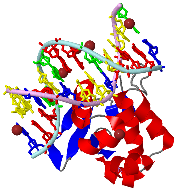
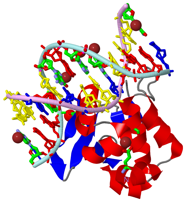
Chain E from PDB  Type:DNA  Length:12
                                            
                 1pp7 E  26 GTTACTTCACTT  37
                                    35  

Chain F from PDB  Type:DNA  Length:13
                                             
                 1pp7 F   3 CAAGTGAAGTAAC  15
                                    12   

Chain U from PDB  Type:PROTEIN  Length:114
 aligned with Q95VR4_TRIVA | Q95VR4 from UniProtKB/TrEMBL  Length:341

    Alignment length:114
                                    14        24        34        44        54        64        74        84        94       104       114    
         Q95VR4_TRIVA     5 DLEASFTSRLPPEIVAALKRKSSRDPNSRFPRKLHMLLTYLASNPQLEEEIGLSWISDTEFKMKKKNVALVMGIKLNTLNVNLRDLAFEQLQHDKGGWTQWKRSGFTRNSVFED 118
               SCOP domains d1pp7u_ U: 39 kda initiator binding protein, IBP39, N-terminal domain                                              SCOP domains
               CATH domains 1pp7U00 U:5-118 'winged helix' repressor DNA binding domain                                                        CATH domains
               Pfam domains IBD-1pp7U01 U:5-112                                                                                         ------ Pfam domains
         Sec.struct. author hhhhhhhh..hhhhhhhhhh........hhhhhhhhhhhhhh.hhhhhhhhheee.....eeehhhhhhhhhh.hhhhhhhhhhhh..eeeeeee..eeeee...ee..ee... Sec.struct. author
                 SAPs(SNPs) ------------------------------------------------------------------------------------------------------------------ SAPs(SNPs)
                    PROSITE ------------------------------------------------------------------------------------------------------------------ PROSITE
                 Transcript ------------------------------------------------------------------------------------------------------------------ Transcript
                 1pp7 U   5 DLEASFTSRLPPEIVAALKRKSSRDPNSRFPRKLHMLLTYLASNPQLEEEIGLSWISDTEFKMKKKNVALVMGIKLNTLNVNLRDLAFEQLQHDKGGWTQWKRSGFTRNSVFED 118
                                    14        24        34        44        54        64        74        84        94       104       114    

   Legend:   → Mismatch (orange background)
  - → Gap (green background, '-', border residues have a numbering label)
    → Modified Residue (blue background, lower-case, 'x' indicates undefined single-letter code, labelled with number + name)
  x → Chemical Group (purple background, 'x', labelled with number + name, e.g. ACE or NH2)
  extra numbering lines below/above indicate numbering irregularities and modified residue names etc., number ends below/above '|'

 Classification and Annotation

(-) SCOP Domains  (1, 1)

Asymmetric/Biological Unit

(-) CATH Domains  (1, 1)

Asymmetric/Biological Unit

(-) Pfam Domains  (1, 1)

Asymmetric/Biological Unit

(-) Gene Ontology  (0, 0)

Asymmetric/Biological Unit(hide GO term definitions)
    (no "Gene Ontology" information available for 1PP7)

 Visualization

(-) Interactive Views

Asymmetric/Biological Unit
  Complete Structure
    Jena3D(integrated viewing of ligand, site, SAP, PROSITE, SCOP information)
    WebMol | AstexViewer[tm]@PDBe
(Java Applets, require no local installation except for Java; loading may be slow)
    STRAP
(Java WebStart application, automatic local installation, requires Java; full application with system access!)
    RasMol
(require local installation)
    Molscript (VRML)
(requires installation of a VRML viewer; select preferred view via VRML and generate a mono or stereo PDF format file)
 
  Ligands, Modified Residues, Ions
    ZN  [ RasMol | Jena3D ]  +environment [ RasMol | Jena3D ]
 
  Sites
    AC1  [ RasMol ]  +environment [ RasMol ]
    AC2  [ RasMol ]  +environment [ RasMol ]
    AC3  [ RasMol ]  +environment [ RasMol ]
    AC4  [ RasMol ]  +environment [ RasMol ]
    AC5  [ RasMol ]  +environment [ RasMol ]
    AC6  [ RasMol ]  +environment [ RasMol ]
    AC7  [ RasMol ]  +environment [ RasMol ]
 
  Cis Peptide Bonds
(no "Cis Peptide Bonds" information available for 1pp7)
 

(-) Still Images

Jmol
  protein: cartoon or spacefill or dots and stick; nucleic acid: cartoon and stick; ligands: spacefill; active site: stick
Molscript
  protein, nucleic acid: cartoon; ligands: spacefill; active site: ball and stick

 Databases and Analysis Tools

(-) Databases

Access by PDB/NDB ID
  1pp7
    Family and Domain InformationProDom | SYSTERS
    General Structural InformationGlycoscienceDB | MMDB | NDB | OCA | PDB | PDBe | PDBj | PDBsum | PDBWiki | PQS | PROTEOPEDIA
    Orientation in MembranesOPM
    Protein SurfaceSURFACE
    Secondary StructureDSSP (structure derived) | HSSP (homology derived)
    Structural GenomicsGeneCensus
    Structural NeighboursCE | VAST
    Structure ClassificationCATH | Dali | SCOP
    Validation and Original DataBMRB Data View | BMRB Restraints Grid | EDS | PROCHECK | RECOORD | WHAT_CHECK
 
Access by UniProt ID/Accession number
  Q95VR4_TRIVA | Q95VR4
    Comparative Protein Structure ModelsModBase
    Genomic InformationEnsembl
    Protein-protein InteractionDIP
    Sequence, Family and Domain InformationInterPro | Pfam | SMART | UniProtKB/TrEMBL
 
Access by Enzyme Classificator   (EC Number)
  (no 'Enzyme Classificator' available)
    General Enzyme InformationBRENDA | EC-PDB | Enzyme | IntEnz
    PathwayKEGG | MetaCyc
 
Access by Disease Identifier   (MIM ID)
  (no 'MIM ID' available)
    Disease InformationOMIM
 
Access by GenAge ID
  (no 'GenAge ID' available)
    Age Related InformationGenAge

(-) Analysis Tools

Access by PDB/NDB ID
    Domain InformationXDom
    Interatomic Contacts of Structural UnitsCSU
    Ligand-protein ContactsLPC
    Protein CavitiescastP
    Sequence and Secondary StructurePDBCartoon
    Structure AlignmentSTRAP(Java WebStart application, automatic local installation, requires Java; full application with system access!)
    Structure and Sequence BrowserSTING
 
Access by UniProt ID/Accession number
  Q95VR4_TRIVA | Q95VR4
    Protein Disorder PredictionDisEMBL | FoldIndex | GLOBPLOT (for more information see DisProt)

 Related Entries

(-) Entries Sharing at Least One Protein Chain (UniProt ID)

UniProtKB/TrEMBL
        Q95VR4_TRIVA | Q95VR41pp8 1q87 1q88 1q89

(-) Related Entries Specified in the PDB File

1pp8 CRYSTAL STRUCTURE OF THE T. VAGINALIS IBP39 INITIATOR BINDING DOMAIN (IBD) BOUND TO THE ALPHA-SCS INR ELEMENT