|
|
|
|
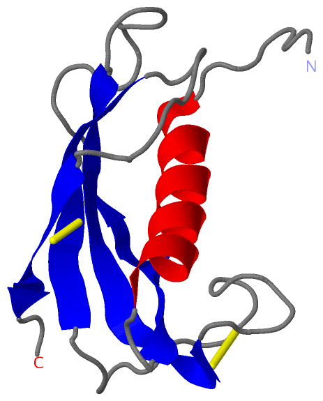
 Description
|
|
Compounds
|
||||||||||||||||||||||||||||||||||||||||||||||||
Chains, Units
Summary Information (see also Sequences/Alignments below) |
Ligands, Modified Residues, Ions (0, 0)| (no "Ligand,Modified Residues,Ions" information available for 1N5H) |
Sites (0, 0)| (no "Site" information available for 1N5H) |
SS Bonds (2, 2)
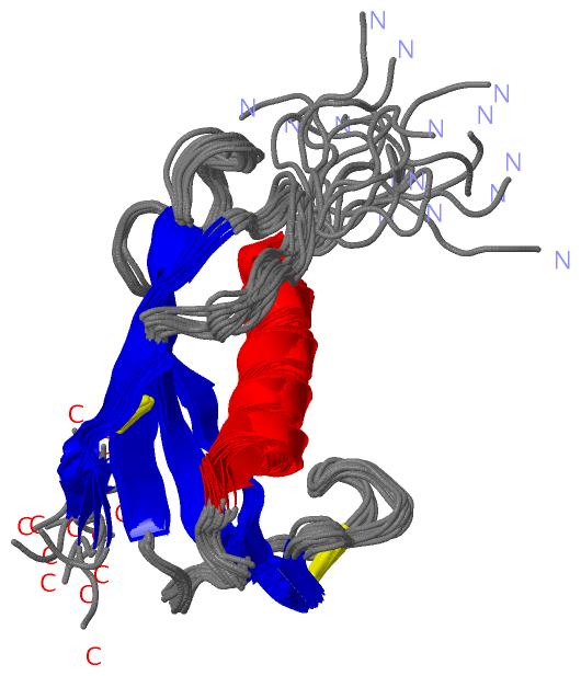
NMR Structure
|
||||||||||||
Cis Peptide Bonds (2, 30)
NMR Structure
|
|||||||||||||||
SAPs(SNPs)/Variants (0, 0)| (no "SAP(SNP)/Variant" information available for 1N5H) |
PROSITE Motifs (2, 2)
NMR Structure (2, 2)
|
||||||||||||||||||||||||||||||||
Exons (0, 0)| (no "Exon" information available for 1N5H) |
Sequences/Alignments
NMR StructureChain A from PDB Type:PROTEIN Length:105 aligned with PG4_PIG | P49933 from UniProtKB/Swiss-Prot Length:149 Alignment length:105 35 45 55 65 75 85 95 105 115 125 PG4_PIG 26 SASAQALSYREAVLRAVDRLNEQSSEANLYRLLELDQPPKADEDPGTPKPVSFTVKETVCPRPTRQPPELCDFKENGRVKQCVGTVTLDQIKDPLDITCNEVQGV 130 SCOP domains d1n5ha_ A: Cathelicidin motif of protegrin-3 SCOP domains CATH domains 1n5hA00 A:26-130 [code=3.10.450.10, no name defined] CATH domains Pfam domains -----Cathelicidins-1n5hA01 A:31-97 --------------------------------- Pfam domains SAPs(SNPs) --------------------------------------------------------------------------------------------------------- SAPs(SNPs) PROSITE --------CATHELICIDINS_------------------------------CATHELICIDINS_2 ------------------------------ PROSITE Transcript --------------------------------------------------------------------------------------------------------- Transcript 1n5h A 26 GSHMQALSYREAVLRAVDRLNEQSSEANLYRLLELDQPPKADEDPGTPKPVSFTVKETVCPRPTRQPPELCDFKENGRVKQCVGTVTLDQIKDPLDITCNEVQGV 130 35 45 55 65 75 85 95 105 115 125
|
||||||||||||||||||||
SCOP Domains (1, 1)
NMR Structure
 |
CATH Domains (1, 1)
NMR Structure
|
Pfam Domains (1, 1)
NMR Structure
|
Gene Ontology (3, 3)|
NMR Structure(hide GO term definitions) Chain A (PG4_PIG | P49933)
|
||||||||||||||||||||||||||||||
Interactive Views
|
||||||||||||||||||||||||||||||||||||||||||||||||||||||||||||||||||||||||||||||||||||||||||||||||||||||||||||||||||||||||||||
Still Images
|
||||||||||||||||
Databases
|
||||||||||||||||||||||||||||||||||||||||||||||||||||||||||||||||||||||||||||||||||||||||||||||||||||||||||||||||||||||||||||||||||||||||||||||||||||||||||||||||
Analysis Tools
|
|||||||||||||||||||||||||||||||||||||||||||||||||||||||||||||
Entries Sharing at Least One Protein Chain (UniProt ID)
Related Entries Specified in the PDB File
|
|