| molecular function |
|---|
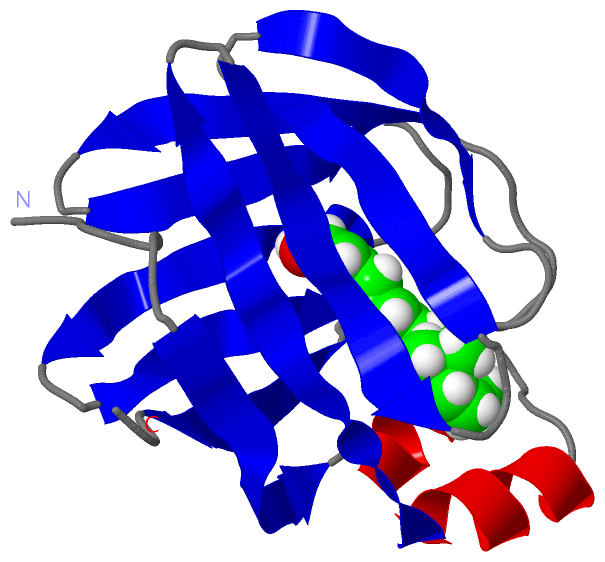
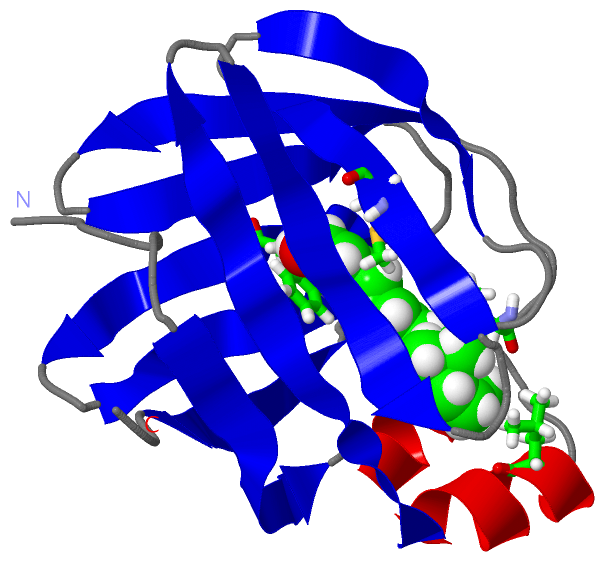
| | GO:0008289 | | lipid binding | | Interacting selectively and non-covalently with a lipid. |
| | GO:0016918 | | retinal binding | | Interacting selectively and non-covalently with retinal, one of the forms of vitamin A. Retinal plays an important role in the visual process in most vertebrates, combining with opsins to form visual pigments in the retina. |
| | GO:0019841 | | retinol binding | | Interacting selectively and non-covalently with retinol, vitamin A1, 2,6,6-trimethyl-1-(9'-hydroxy-3',7'-dimethylnona-1',3',5',7'-tetraenyl)cyclohex-1-ene, one of the three components that makes up vitamin A. Retinol is an intermediate in the vision cycle and it also plays a role in growth and differentiation. |
| | GO:0005215 | | transporter activity | | Enables the directed movement of substances (such as macromolecules, small molecules, ions) into, out of or within a cell, or between cells. |
| biological process |
|---|
| | GO:0030852 | | regulation of granulocyte differentiation | | Any process that modulates the frequency, rate or extent of granulocyte differentiation. |
| | GO:0033189 | | response to vitamin A | | Any process that results in a change in state or activity of a cell or an organism (in terms of movement, secretion, enzyme production, gene expression, etc.) as a result of a vitamin A stimulus. |
| | GO:0002138 | | retinoic acid biosynthetic process | | The chemical reactions and pathways resulting in the biosynthesis of retinoic acid, one of the three components that makes up vitamin A. |
| | GO:0042573 | | retinoic acid metabolic process | | The chemical reactions and pathways involving retinoic acid, one of the three components that makes up vitamin A. |
| | GO:0042572 | | retinol metabolic process | | The chemical reactions and pathways involving retinol, one of the three compounds that makes up vitamin A. |
| | GO:0006810 | | transport | | The directed movement of substances (such as macromolecules, small molecules, ions) or cellular components (such as complexes and organelles) into, out of or within a cell, or between cells, or within a multicellular organism by means of some agent such as a transporter, pore or motor protein. |
| | GO:0006776 | | vitamin A metabolic process | | The chemical reactions and pathways involving any of the vitamin A compounds, retinol, retinal (retinaldehyde) and retinoic acid, all of which are derivatives of beta-carotene. |
| cellular component |
|---|
| | GO:0044297 | | cell body | | The portion of a cell bearing surface projections such as axons, dendrites, cilia, or flagella that includes the nucleus, but excludes all cell projections. |
| | GO:0005737 | | cytoplasm | | All of the contents of a cell excluding the plasma membrane and nucleus, but including other subcellular structures. |
| | GO:0005829 | | cytosol | | The part of the cytoplasm that does not contain organelles but which does contain other particulate matter, such as protein complexes. |
 Description