|
|
|
|
 Description
|
|
Compounds
|
||||||||||||||||||||||||||||||||||||||||||||
Chains, Units
Summary Information (see also Sequences/Alignments below) |
Ligands, Modified Residues, Ions (0, 0)| (no "Ligand,Modified Residues,Ions" information available for 1MI0) |
Sites (0, 0)| (no "Site" information available for 1MI0) |
SS Bonds (0, 0)| (no "SS Bond" information available for 1MI0) |
Cis Peptide Bonds (0, 0)| (no "Cis Peptide Bond" information available for 1MI0) |
SAPs(SNPs)/Variants (0, 0)| (no "SAP(SNP)/Variant" information available for 1MI0) |
PROSITE Motifs (0, 0)| (no "PROSITE Motif" information available for 1MI0) |
Exons (0, 0)| (no "Exon" information available for 1MI0) |
Sequences/Alignments
Asymmetric UnitChain A from PDB Type:PROTEIN Length:61 aligned with Q53291_FINMA | Q53291 from UniProtKB/TrEMBL Length:455 Alignment length:63 334 333 | 333 | 341 351 361 371 381 Q53291_FINMA 324 PEEPMDTYKL--ILNGKTLKGETTTEAVDAATAEKVFKQYANDNGVDGEWTYDDATKTFTVTE 384 SCOP domains d1mi0a_ A: SCOP domains CATH domains 1mi0A00 A:1-61 [cod e=3.10.20.10, no name defined] CATH domains Pfam domains --------------------------------------------------------------- Pfam domains SAPs(SNPs) --------------------------------------------------------------- SAPs(SNPs) PROSITE --------------------------------------------------------------- PROSITE Transcript --------------------------------------------------------------- Transcript 1mi0 A 1 HHHAMDTYKLVIVLNGTTFT--YTTEAVDAATAEKVFKQYANDNGVDGEWTYADATKTFTVTE 61 10 20 | 28 38 48 58 20 21 Chain B from PDB Type:PROTEIN Length:62 aligned with Q53291_FINMA | Q53291 from UniProtKB/TrEMBL Length:455 Alignment length:64 334 333 | 332| | 340 350 360 370 380 Q53291_FINMA 323 KPEEPMDTYKL--ILNGKTLKGETTTEAVDAATAEKVFKQYANDNGVDGEWTYDDATKTFTVTE 384 SCOP domains d1mi0b_ B: SCOP domains CATH domains 1mi0B00 B:1-62 [code =3.10.20.10, no name defined] CATH domains Pfam domains ---------------------------------------------------------------- Pfam domains SAPs(SNPs) ---------------------------------------------------------------- SAPs(SNPs) PROSITE ---------------------------------------------------------------- PROSITE Transcript ---------------------------------------------------------------- Transcript 1mi0 B 1 HHHHAMDTYKLVIVLNGTTFT--YTTEAVDAATAEKVFKQYANDNGVDGEWTYADATKTFTVTE 62 10 20| | 28 38 48 58 21 22
|
||||||||||||||||||||
SCOP Domains (1, 2)
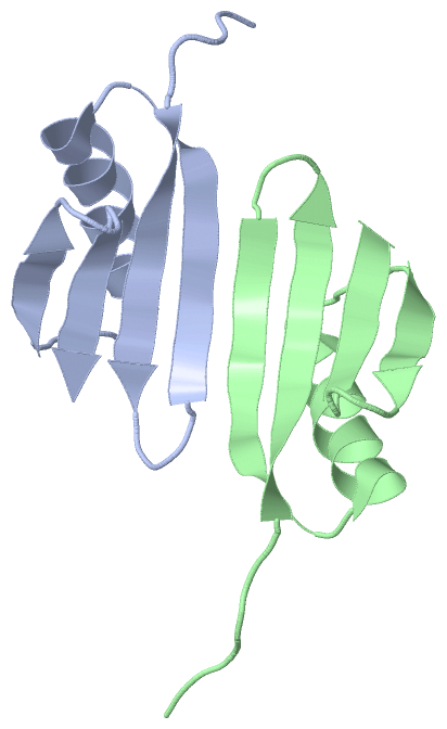
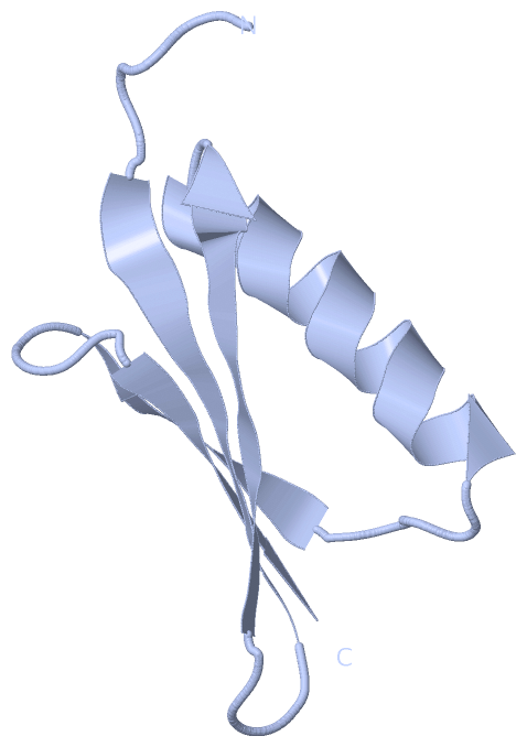
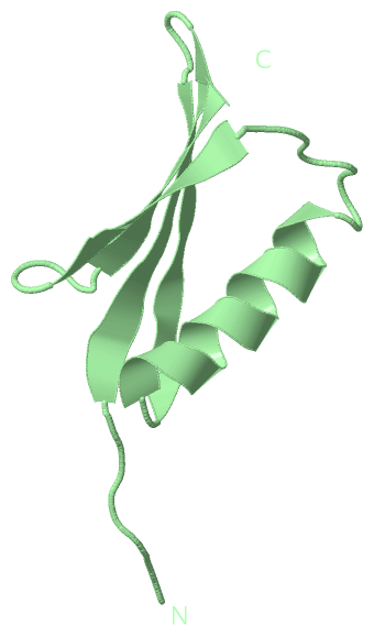
Asymmetric Unit
 |
CATH Domains (1, 2)
Asymmetric Unit
|
Pfam Domains (0, 0)| (no "Pfam Domain" information available for 1MI0) |
Gene Ontology (1, 1)|
Asymmetric Unit(hide GO term definitions) Chain A,B (Q53291_FINMA | Q53291)
|
||||||||||||
Interactive Views
|
|||||||||||||||||||||||||||||||||||||||||||||||||||||||||||||||||||||||||||||||||||||||||||||||||||||||||||||||||||||||||||||||||||||||||||
Still Images
|
||||||||||||||||
Databases
|
||||||||||||||||||||||||||||||||||||||||||||||||||||||||||||||||||||||||||||||||||||||||||||||||||||||||||||||||||||||||||||||||||||||||||||||||||||||||||||||||
Analysis Tools
|
|||||||||||||||||||||||||||||||||||||||||||||||||||||||||||||
Entries Sharing at Least One Protein Chain (UniProt ID)
Related Entries Specified in the PDB File
|
|