Show PDB file:   
         Plain Text   HTML   (compressed file size)
QuickSearch:   
by PDB,NDB,UniProt,PROSITE Code or Search Term(s)  
(-)NMR Structure - manually
(-)NMR Structure - model 1
(-)NMR Structure - all models
collapse expand < >
Image NMR Structure - manually
NMR Structure - manually  (Jmol Viewer)
Image NMR Structure - model 1
NMR Structure - model 1  (Jmol Viewer)
Image NMR Structure - all models
NMR Structure - all models  (Jmol Viewer)

(-) Description

Title :  REGULATION OF DIRECTIONALITY IN BACTERIOPHAGE LAMBDA SITE-SPECIFIC RECOMBINATION: STRUCTURE OF THE XIS PROTEIN
 
Authors :  M. D. Sam, C. Papagiannis, K. M. Connolly, L. Corselli, J. Iwahara, J. Lee, M. Phillips, J. M. Wojciak, R. C. Johnson, R. T. Clubb
Date :  04 Jun 02  (Deposition) - 10 Jun 03  (Release) - 24 Feb 09  (Revision)
Method :  SOLUTION NMR
Resolution :  NOT APPLICABLE
Chains :  NMR Structure  :  A  (20x)
Keywords :  Dna Architectural Protein, 'Winged'-Helix Protein, Phage Excision, Site-Specific Dna Recombination, Viral Protein (Keyword Search: [Gene Ontology, PubMed, Web (Google))
 
Reference :  M. D. Sam, C. Papagiannis, K. M. Connolly, L. Corselli, J. Iwahara, J. Lee, M. Phillips, J. M. Wojciak, R. C. Johnson, R. T. Clubb
Regulation Of Directionality In Bacteriophage Lambda Site-Specific Recombination: Structure Of The Xis Protein
J. Mol. Biol. V. 324 791 2002
PubMed-ID: 12460578  |  Reference-DOI: 10.1016/S0022-2836(02)01150-6
(for further references see the PDB file header)

(-) Compounds

Molecule 1 - EXCISIONASE
    ChainsA
    EngineeredYES
    Expression SystemESCHERICHIA COLI
    Expression System PlasmidPBAD33
    Expression System StrainRJ3386 (BL21-DE3)
    Expression System Taxid562
    Expression System Vector TypePLASMID
    FragmentDNA BINDING DOMAIN
    GeneXIS
    MutationYES
    Organism ScientificENTEROBACTERIA PHAGE LAMBDA
    Organism Taxid10710
    SynonymXIS

 Structural Features

(-) Chains, Units

  
NMR Structure (20x)

Summary Information (see also Sequences/Alignments below)

(-) Ligands, Modified Residues, Ions  (0, 0)

(no "Ligand,Modified Residues,Ions" information available for 1LX8)

(-) Sites  (0, 0)

(no "Site" information available for 1LX8)

(-) SS Bonds  (0, 0)

(no "SS Bond" information available for 1LX8)

(-) Cis Peptide Bonds  (1, 20)
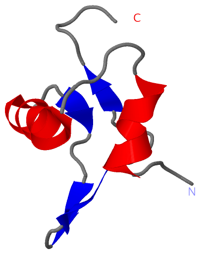
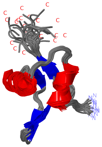
NMR Structure
No.ModelResidues
11, 2, 3, 4, 5, 6, 7, 8, 9, 10, 11, 12, 13, 14, 15, 16, 17, 18, 19, 20Phe A:31 -Pro A:32

 Sequence-Structure Mapping

(-) SAPs(SNPs)/Variants  (0, 0)

(no "SAP(SNP)/Variant" information available for 1LX8)

(-) PROSITE Motifs  (0, 0)

(no "PROSITE Motif" information available for 1LX8)

(-) Exons   (0, 0)

(no "Exon" information available for 1LX8)

(-) Sequences/Alignments

NMR Structure
   Reformat: Number of residues per line =  ('0' or empty: single-line sequence representation)
  Number of residues per labelling interval =   
  UniProt sequence: complete  aligned part    
   Show mapping: SCOP domains CATH domains Pfam domains Secondary structure (by author)
SAPs(SNPs) PROSITE motifs Exons
(details for a mapped element are shown in a popup box when the mouse pointer rests over it)
Chain A from PDB  Type:PROTEIN  Length:55
 aligned with VXIS_LAMBD | P03699 from UniProtKB/Swiss-Prot  Length:72

    Alignment length:55
                                    10        20        30        40        50     
            VXIS_LAMBD    1 MYLTLQEWNARQRRPRSLETVRRWVRECRIFPPPVKDGREYLFHESAVKVDLNRP 55
               SCOP domains d1lx8a_ A: Excisionase Xis                              SCOP domains
               CATH domains 1lx8A00 A:1-55  [code=1.10.1660.20, no name defined]    CATH domains
               Pfam domains Exc-1lx8A01 A:1-55                                      Pfam domains
         Sec.struct. author ...hhhhhhhh.....hhhhhhhhhhh..ee...eee..eee.....ee...... Sec.struct. author
                 SAPs(SNPs) ------------------------------------------------------- SAPs(SNPs)
                    PROSITE ------------------------------------------------------- PROSITE
                 Transcript ------------------------------------------------------- Transcript
                  1lx8 A  1 MYLTLQEWNARQRRPRSLETVRRWVRESRIFPPPVKDGREYLFHESAVKVDLNRP 55
                                    10        20        30        40        50     

   Legend:   → Mismatch (orange background)
  - → Gap (green background, '-', border residues have a numbering label)
    → Modified Residue (blue background, lower-case, 'x' indicates undefined single-letter code, labelled with number + name)
  x → Chemical Group (purple background, 'x', labelled with number + name, e.g. ACE or NH2)
  extra numbering lines below/above indicate numbering irregularities and modified residue names etc., number ends below/above '|'

 Classification and Annotation

(-) SCOP Domains  (1, 1)

NMR Structure

(-) CATH Domains  (1, 1)

NMR Structure

(-) Pfam Domains  (1, 1)

NMR Structure
(-)
Clan: HTH (544)

(-) Gene Ontology  (2, 2)

NMR Structure(hide GO term definitions)
Chain A   (VXIS_LAMBD | P03699)
molecular function
    GO:0003677    DNA binding    Any molecular function by which a gene product interacts selectively and non-covalently with DNA (deoxyribonucleic acid).
biological process
    GO:0006310    DNA recombination    Any process in which a new genotype is formed by reassortment of genes resulting in gene combinations different from those that were present in the parents. In eukaryotes genetic recombination can occur by chromosome assortment, intrachromosomal recombination, or nonreciprocal interchromosomal recombination. Intrachromosomal recombination occurs by crossing over. In bacteria it may occur by genetic transformation, conjugation, transduction, or F-duction.

 Visualization

(-) Interactive Views

NMR Structure
  Complete Structure
    Jena3D(integrated viewing of ligand, site, SAP, PROSITE, SCOP information)
    WebMol | AstexViewer[tm]@PDBe
(Java Applets, require no local installation except for Java; loading may be slow)
    STRAP
(Java WebStart application, automatic local installation, requires Java; full application with system access!)
    RasMol
(require local installation)
    Molscript (VRML)
(requires installation of a VRML viewer; select preferred view via VRML and generate a mono or stereo PDF format file)
 
  Ligands, Modified Residues, Ions
(no "Ligands, Modified Residues, Ions" information available for 1lx8)
 
  Sites
(no "Sites" information available for 1lx8)
 
  Cis Peptide Bonds
    Phe A:31 - Pro A:32   [ RasMol ]  
 

(-) Still Images

Jmol
  protein: cartoon or spacefill or dots and stick; nucleic acid: cartoon and stick; ligands: spacefill; active site: stick
Molscript
  protein, nucleic acid: cartoon; ligands: spacefill; active site: ball and stick

 Databases and Analysis Tools

(-) Databases

Access by PDB/NDB ID
  1lx8
    Family and Domain InformationProDom | SYSTERS
    General Structural InformationGlycoscienceDB | MMDB | NDB | OCA | PDB | PDBe | PDBj | PDBsum | PDBWiki | PQS | PROTEOPEDIA
    Orientation in MembranesOPM
    Protein SurfaceSURFACE
    Secondary StructureDSSP (structure derived) | HSSP (homology derived)
    Structural GenomicsGeneCensus
    Structural NeighboursCE | VAST
    Structure ClassificationCATH | Dali | SCOP
    Validation and Original DataBMRB Data View | BMRB Restraints Grid | EDS | PROCHECK | RECOORD | WHAT_CHECK
 
Access by UniProt ID/Accession number
  VXIS_LAMBD | P03699
    Comparative Protein Structure ModelsModBase
    Genomic InformationEnsembl
    Protein-protein InteractionDIP
    Sequence, Family and Domain InformationInterPro | Pfam | SMART | UniProtKB/SwissProt
 
Access by Enzyme Classificator   (EC Number)
  (no 'Enzyme Classificator' available)
    General Enzyme InformationBRENDA | EC-PDB | Enzyme | IntEnz
    PathwayKEGG | MetaCyc
 
Access by Disease Identifier   (MIM ID)
  (no 'MIM ID' available)
    Disease InformationOMIM
 
Access by GenAge ID
  (no 'GenAge ID' available)
    Age Related InformationGenAge

(-) Analysis Tools

Access by PDB/NDB ID
    Domain InformationXDom
    Interatomic Contacts of Structural UnitsCSU
    Ligand-protein ContactsLPC
    Protein CavitiescastP
    Sequence and Secondary StructurePDBCartoon
    Structure AlignmentSTRAP(Java WebStart application, automatic local installation, requires Java; full application with system access!)
    Structure and Sequence BrowserSTING
 
Access by UniProt ID/Accession number
  VXIS_LAMBD | P03699
    Protein Disorder PredictionDisEMBL | FoldIndex | GLOBPLOT (for more information see DisProt)

 Related Entries

(-) Entries Sharing at Least One Protein Chain (UniProt ID)

UniProtKB/Swiss-Prot
        VXIS_LAMBD | P036991rh6 2ief 2og0 5j0n

(-) Related Entries Specified in the PDB File

(no "Related Entries Specified in the PDB File" available for 1LX8)