|
|
|
|
 Description
|
|
Compounds
|
||||||||||||||||||||||||||||||||||||||||||||||||||||
Chains, Units
Summary Information (see also Sequences/Alignments below) |
Ligands, Modified Residues, Ions (5, 12)
Asymmetric Unit (5, 12)
|
Sites (12, 12)
Asymmetric Unit (12, 12)
|
SS Bonds (10, 10)
Asymmetric Unit
|
||||||||||||||||||||||||||||||||||||||||||||
Cis Peptide Bonds (2, 2)
Asymmetric Unit
|
||||||||||||
SAPs(SNPs)/Variants (0, 0)| (no "SAP(SNP)/Variant" information available for 1LCS) |
PROSITE Motifs (0, 0)| (no "PROSITE Motif" information available for 1LCS) |
Exons (0, 0)| (no "Exon" information available for 1LCS) |
Sequences/Alignments
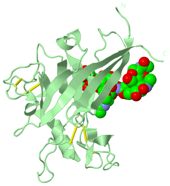
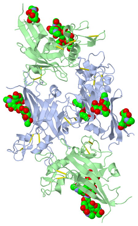
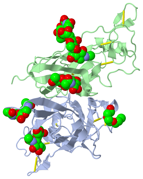
Asymmetric UnitChain A from PDB Type:PROTEIN Length:205 aligned with ENV_FLVLB | P11261 from UniProtKB/Swiss-Prot Length:662 Alignment length:205 47 57 67 77 87 97 107 117 127 137 147 157 167 177 187 197 207 217 227 237 ENV_FLVLB 38 PHQIYNVTWTITNLVTGTKANATSMLGTLTDAFPTMYFDLCDIIGNTWNPSDQEPFPGYGCDQPMRRWQQRNTPFYVCPGHANRKQCGGPQDGFCAVWGCETTGETYWRPTSSWDYITVKKGVTQGIYQCSGGGWCGPCYDKAVHSSITGASEGGRCNPLILQFTQKGRQTSWDGPKSWGLRLYRSGYDPIALFSVSRQVMTITL 242 SCOP domains d1lcsa_ A: FLV receptor-binding domain SCOP domains CATH domains 1lcsA00 A:4-208 Viral Glycoprotein Gp70 CATH domains Pfam domains ------------------------------------------------------------------------------------------------------------------------------------------------------------------------------------------------------------- Pfam domains SAPs(SNPs) ------------------------------------------------------------------------------------------------------------------------------------------------------------------------------------------------------------- SAPs(SNPs) PROSITE ------------------------------------------------------------------------------------------------------------------------------------------------------------------------------------------------------------- PROSITE Transcript ------------------------------------------------------------------------------------------------------------------------------------------------------------------------------------------------------------- Transcript 1lcs A 4 PHQVYNVTWTITNLVTGTKANATSMLGTLTDAFPTMYFDLCDIIGNTWNPSDQEPFPGYGCDQPMRRWQQRNTPFYVCPGHANRKQCGGPQDGFCAVWGCETTGETYWRPTSSWDYITVKKGVTQGIYQCSGGGWCGPCYDKAVHSSTTGASEGGRCNPLILQFTQKGRQTSWDGPKSWGLRLYRSGYDPIALFSVSRQVMTITP 208 13 23 33 43 53 63 73 83 93 103 113 123 133 143 153 163 173 183 193 203 Chain B from PDB Type:PROTEIN Length:205 aligned with ENV_FLVLB | P11261 from UniProtKB/Swiss-Prot Length:662 Alignment length:205 47 57 67 77 87 97 107 117 127 137 147 157 167 177 187 197 207 217 227 237 ENV_FLVLB 38 PHQIYNVTWTITNLVTGTKANATSMLGTLTDAFPTMYFDLCDIIGNTWNPSDQEPFPGYGCDQPMRRWQQRNTPFYVCPGHANRKQCGGPQDGFCAVWGCETTGETYWRPTSSWDYITVKKGVTQGIYQCSGGGWCGPCYDKAVHSSITGASEGGRCNPLILQFTQKGRQTSWDGPKSWGLRLYRSGYDPIALFSVSRQVMTITL 242 SCOP domains d1lcsb_ B: FLV receptor-binding domain SCOP domains CATH domains 1lcsB00 B:4-208 Viral Glycoprotein Gp70 CATH domains Pfam domains (1) TLV_coat-1lcsB01 B:4-208 Pfam domains (1) Pfam domains (2) TLV_coat-1lcsB02 B:4-208 Pfam domains (2) SAPs(SNPs) ------------------------------------------------------------------------------------------------------------------------------------------------------------------------------------------------------------- SAPs(SNPs) PROSITE ------------------------------------------------------------------------------------------------------------------------------------------------------------------------------------------------------------- PROSITE Transcript ------------------------------------------------------------------------------------------------------------------------------------------------------------------------------------------------------------- Transcript 1lcs B 4 PHQVYNVTWTITNLVTGTKANATSMLGTLTDAFPTMYFDLCDIIGNTWNPSDQEPFPGYGCDQPMRRWQQRNTPFYVCPGHANRKQCGGPQDGFCAVWGCETTGETYWRPTSSWDYITVKKGVTQGIYQCSGGGWCGPCYDKAVHSSTTGASEGGRCNPLILQFTQKGRQTSWDGPKSWGLRLYRSGYDPIALFSVSRQVMTITP 208 13 23 33 43 53 63 73 83 93 103 113 123 133 143 153 163 173 183 193 203
|
||||||||||||||||||||
SCOP Domains (1, 2)
Asymmetric Unit
 |
CATH Domains (1, 2)
Asymmetric Unit
|
Pfam Domains (1, 2)
Asymmetric Unit
|
Gene Ontology (15, 15)|
Asymmetric Unit(hide GO term definitions) Chain A,B (ENV_FLVLB | P11261)
|
||||||||||||||||||||||||||||||||||||||||||||||||||||||||||||||||||||||||||||||||||||||||||||||||||||||||||||
Interactive Views
|
||||||||||||||||||||||||||||||||||||||||||||||||||||||||||||||||||||||||||||||||||||||||||||||||||||||||||||||||||||||||||||||||||||||||||||||||||||||||||||||||||||||||||||||||||||||||||||||||||||||||||||||||||||||||||||||||||||||||||||||||||||||||||||||||||||||||
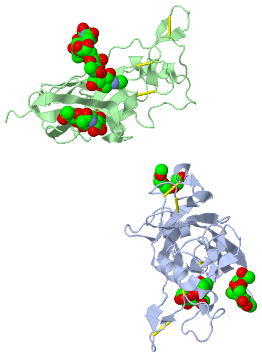
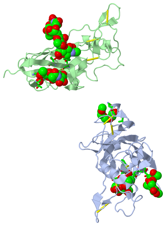
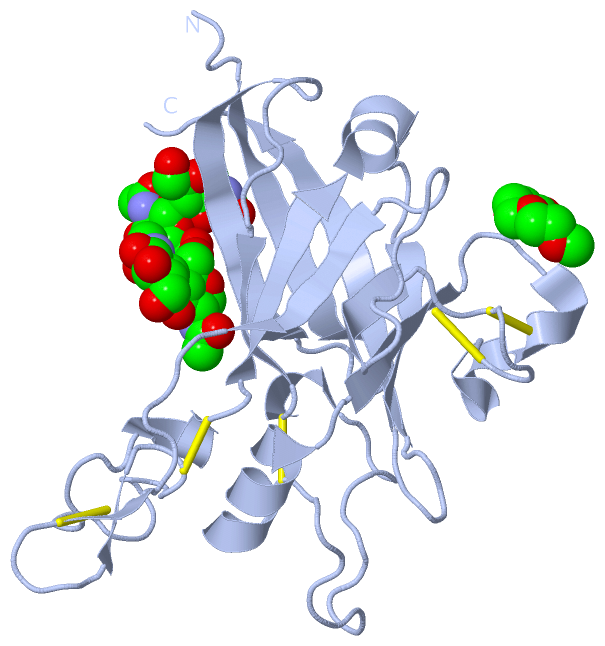
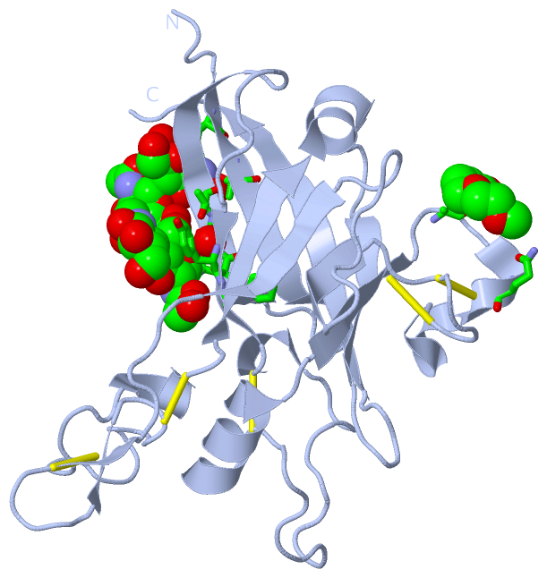
Still Images
|
||||||||||||||||
Databases
|
||||||||||||||||||||||||||||||||||||||||||||||||||||||||||||||||||||||||||||||||||||||||||||||||||||||||||||||||||||||||||||||||||||||||||||||||||||||||||||||||
Analysis Tools
|
|||||||||||||||||||||||||||||||||||||||||||||||||||||||||||||
Entries Sharing at Least One Protein Chain (UniProt ID)
Related Entries Specified in the PDB File
|
|