|
|
|
|
 Description
|
|
Compounds
|
||||||||||||||||||||||||||||||||||||||||||||||||
Chains, Units
Summary Information (see also Sequences/Alignments below) |
Ligands, Modified Residues, Ions (0, 0)| (no "Ligand,Modified Residues,Ions" information available for 1IJA) |
Sites (0, 0)| (no "Site" information available for 1IJA) |
SS Bonds (0, 0)| (no "SS Bond" information available for 1IJA) |
Cis Peptide Bonds (0, 0)| (no "Cis Peptide Bond" information available for 1IJA) |
SAPs(SNPs)/Variants (0, 0)| (no "SAP(SNP)/Variant" information available for 1IJA) |
PROSITE Motifs (0, 0)| (no "PROSITE Motif" information available for 1IJA) |
Exons (0, 0)| (no "Exon" information available for 1IJA) |
Sequences/Alignments
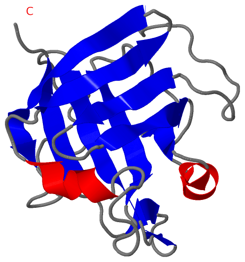
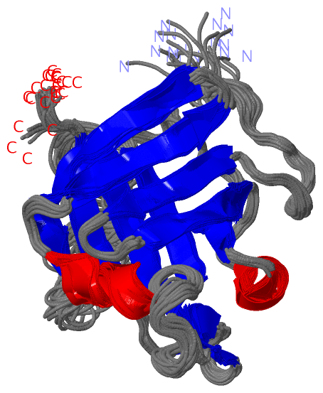
NMR StructureChain A from PDB Type:PROTEIN Length:148 aligned with Q9S446_STAAU | Q9S446 from UniProtKB/TrEMBL Length:206 Alignment length:148 68 78 88 98 108 118 128 138 148 158 168 178 188 198 Q9S446_STAAU 59 QQAKPQIPKDKSKVAGYIEIPDADIKEPVYPGPATPEQLNRGVSFAEENESLDDQNISIAGHTFIDRPNYQFTNLKAAKKGSMVYFKVGNETRKYKMTSIRDVKPTDVGVLDEQKGKDKQLTLITCDDYNEKTGVWEKRKIFVATEVK 206 SCOP domains d1ijaa_ A: Sortase A SCOP domains CATH domains 1ijaA00 A:1-148 [code=2.40.260.10, no name defined] CATH domains Pfam domains ---------------------------------------------------------------------------------------------------------------------------------------------------- Pfam domains SAPs(SNPs) ---------------------------------------------------------------------------------------------------------------------------------------------------- SAPs(SNPs) PROSITE ---------------------------------------------------------------------------------------------------------------------------------------------------- PROSITE Transcript ---------------------------------------------------------------------------------------------------------------------------------------------------- Transcript 1ija A 1 MQAKPQIPKDKSKVAGYIEIPDADIKEPVYPGPATPEQLNRGVSFAEENESLDDQNISIAGHTFIDRPNYQFTNLKAAKKGSMVYFKVGNETRKYKMTSIRDVKPTDVGVLDEQKGKDKQLTLITCDDYNEKTGVWEKRKIFVATEVK 148 10 20 30 40 50 60 70 80 90 100 110 120 130 140
|
||||||||||||||||||||
SCOP Domains (1, 1)
NMR Structure
 |
CATH Domains (1, 1)
NMR Structure
|
Pfam Domains (0, 0)| (no "Pfam Domain" information available for 1IJA) |
Gene Ontology (1, 1)|
NMR Structure(hide GO term definitions) Chain A (Q9S446_STAAU | Q9S446)
|
||||||||||||
Interactive Views
|
||||||||||||||||||||||||||||||||||||||||||||||||||||||||||||||||||||||||||||||||||||||||||||||||||||||||||||||||||||
Still Images
|
||||||||||||||||
Databases
|
||||||||||||||||||||||||||||||||||||||||||||||||||||||||||||||||||||||||||||||||||||||||||||||||||||||||||||||||||||||||||||||||||||||||||||||||||||||||||||||||
Analysis Tools
|
|||||||||||||||||||||||||||||||||||||||||||||||||||||||||||||
Entries Sharing at Least One Protein Chain (UniProt ID)
Related Entries Specified in the PDB File
|
|