|
|
|
|
 Description
|
|
Compounds
|
||||||||||||||||||||||||||||||||||||||||||||||||||||||||||||||||||||||
Chains, Units
Summary Information (see also Sequences/Alignments below) |
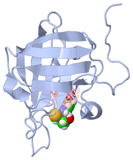
Ligands, Modified Residues, Ions (3, 3)| NMR Structure (3, 3) NMR Structure * (2, 2) |
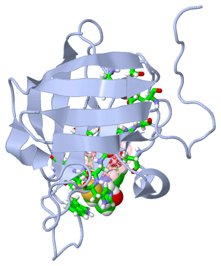
Sites (2, 2)
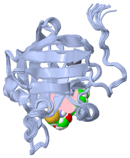
NMR Structure (2, 2)
|
SS Bonds (0, 0)| (no "SS Bond" information available for 2KID) |
Cis Peptide Bonds (1, 20)
NMR Structure
|
||||||||||
SAPs(SNPs)/Variants (0, 0)| (no "SAP(SNP)/Variant" information available for 2KID) |
PROSITE Motifs (0, 0)| (no "PROSITE Motif" information available for 2KID) |
Exons (0, 0)| (no "Exon" information available for 2KID) |
Sequences/Alignments
NMR StructureChain A from PDB Type:PROTEIN Length:148 aligned with Q9S446_STAAU | Q9S446 from UniProtKB/TrEMBL Length:206 Alignment length:148 68 78 88 98 108 118 128 138 148 158 168 178 188 198 Q9S446_STAAU 59 QQAKPQIPKDKSKVAGYIEIPDADIKEPVYPGPATPEQLNRGVSFAEENESLDDQNISIAGHTFIDRPNYQFTNLKAAKKGSMVYFKVGNETRKYKMTSIRDVKPTDVGVLDEQKGKDKQLTLITCDDYNEKTGVWEKRKIFVATEVK 206 SCOP domains ---------------------------------------------------------------------------------------------------------------------------------------------------- SCOP domains CATH domains 2kidA00 A:59-206 [code=2.40.260.10, no name defined] CATH domains Pfam domains -----------------Sortase-2kidA01 A:76-205 - Pfam domains SAPs(SNPs) ---------------------------------------------------------------------------------------------------------------------------------------------------- SAPs(SNPs) PROSITE ---------------------------------------------------------------------------------------------------------------------------------------------------- PROSITE Transcript ---------------------------------------------------------------------------------------------------------------------------------------------------- Transcript 2kid A 59 MQAKPQIPKDKSKVAGYIEIPDADIKEPVYPGPATPEQLNRGVSFAEENESLDDQNISIAGHTFIDRPNYQFTNLKAAKKGSMVYFKVGNETRKYKMTSIRDVKPTDVGVLDEQKGKDKQLTLITCDDYNEKTGVWEKRKIFVATEVK 206 68 78 88 98 108 118 128 138 148 158 168 178 188 198
Chain C from PDB Type:PROTEIN Length:5
SCOP domains ----- SCOP domains
CATH domains ----- CATH domains
Pfam domains ----- Pfam domains
SAPs(SNPs) ----- SAPs(SNPs)
PROSITE ----- PROSITE
Transcript ----- Transcript
2kid C 701 xLPAt 705
| |
701-PHQ
705-B27
|
||||||||||||||||||||
SCOP Domains (0, 0)| (no "SCOP Domain" information available for 2KID) |
CATH Domains (1, 1)
NMR Structure
 |
Pfam Domains (1, 1)
NMR Structure
|
Gene Ontology (1, 1)|
NMR Structure(hide GO term definitions) Chain A (Q9S446_STAAU | Q9S446)
|
||||||||||||
Interactive Views
|
||||||||||||||||||||||||||||||||||||||||||||||||||||||||||||||||||||||||||||||||||||||||||||||||||||||||||||||||||||||||||||||||||||||||||||
Still Images
|
||||||||||||||||
Databases
|
||||||||||||||||||||||||||||||||||||||||||||||||||||||||||||||||||||||||||||||||||||||||||||||||||||||||||||||||||||||||||||||||||||||||||||||||||||||||||||||||
Analysis Tools
|
|||||||||||||||||||||||||||||||||||||||||||||||||||||||||||||
Entries Sharing at Least One Protein Chain (UniProt ID)
Related Entries Specified in the PDB File
|
|