|
|
|
|
 Description
|
|
Compounds
|
||||||||||||||||||||||||||||||||||||||||||||||||
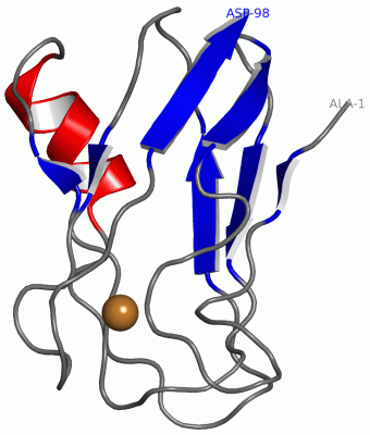
Chains, Units
Summary Information (see also Sequences/Alignments below) |
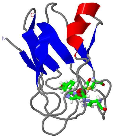
Ligands, Modified Residues, Ions (1, 1)
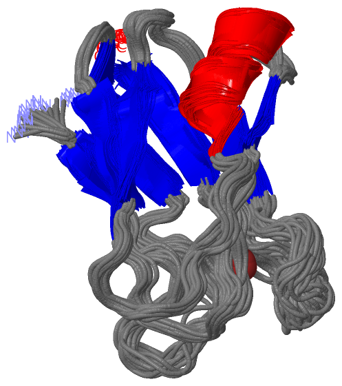
NMR Structure (1, 1)
|
Sites (1, 1)
NMR Structure (1, 1)
|
SS Bonds (0, 0)| (no "SS Bond" information available for 1J5C) |
Cis Peptide Bonds (2, 70)
NMR Structure
|
|||||||||||||||
SAPs(SNPs)/Variants (0, 0)| (no "SAP(SNP)/Variant" information available for 1J5C) |
PROSITE Motifs (1, 1)
NMR Structure (1, 1)
|
||||||||||||||||||||||||
Exons (0, 0)| (no "Exon" information available for 1J5C) |
Sequences/Alignments
NMR StructureChain A from PDB Type:PROTEIN Length:98 aligned with PLAS_SYNY3 | P21697 from UniProtKB/Swiss-Prot Length:126 Alignment length:98 38 48 58 68 78 88 98 108 118 PLAS_SYNY3 29 ANATVKMGSDSGALVFEPSTVTIKAGEEVKWVNNKLSPHNIVFAADGVDADTAAKLSHKGLAFAAGESFTSTFTEPGTYTYYCEPHRGAGMVGKVVVE 126 SCOP domains d1j5ca_ A: Plastocyanin SCOP domains CATH domains 1j5cA00 A:1-98 Cupredoxins - blue copper proteins CATH domains Pfam domains -------------------------------------------------------------------------------------------------- Pfam domains SAPs(SNPs) -------------------------------------------------------------------------------------------------- SAPs(SNPs) PROSITE ----------------------------------------------------------------------------COPPER_BLUE ------- PROSITE Transcript -------------------------------------------------------------------------------------------------- Transcript 1j5c A 1 ANATVKMGSDSGALVFEPSTVTIKAGEEVKWVNNKLSPHNIVFAADGVDADTAAKLSHKGLAFAAGESFTSTFTEPGTYTYYCEPHRGAGMVGKVVVD 98 10 20 30 40 50 60 70 80 90
|
||||||||||||||||||||
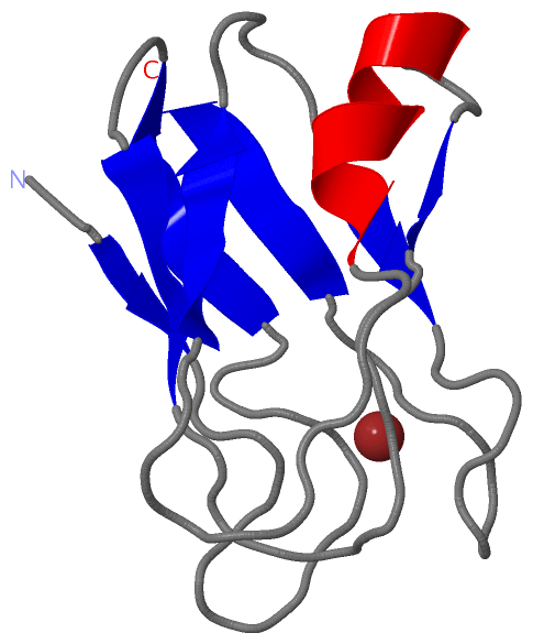
SCOP Domains (1, 1)
NMR Structure
 |
CATH Domains (1, 1)
NMR Structure
|
Pfam Domains (0, 0)| (no "Pfam Domain" information available for 1J5C) |
Gene Ontology (7, 7)|
NMR Structure(hide GO term definitions) Chain A (PLAS_SYNY3 | P21697)
|
||||||||||||||||||||||||||||||||||||||||||||||||||||||||||||
Interactive Views
|
||||||||||||||||||||||||||||||||||||||||||||||||||||||||||||||||||||||||||||||||||||||||||||||||||||||||||||||||||||||||||||||
Still Images
|
||||||||||||||||
Databases
|
||||||||||||||||||||||||||||||||||||||||||||||||||||||||||||||||||||||||||||||||||||||||||||||||||||||||||||||||||||||||||||||||||||||||||||||||||||||||||||||||
Analysis Tools
|
|||||||||||||||||||||||||||||||||||||||||||||||||||||||||||||
Entries Sharing at Least One Protein Chain (UniProt ID)
Related Entries Specified in the PDB File
|
|