|
|
|
|
 Description
|
|
Compounds
|
||||||||||||||||||||||||||||
Chains, Units
Summary Information (see also Sequences/Alignments below) |
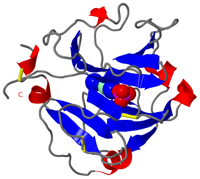
Ligands, Modified Residues, Ions (3, 4)| Asymmetric/Biological Unit (3, 4) |
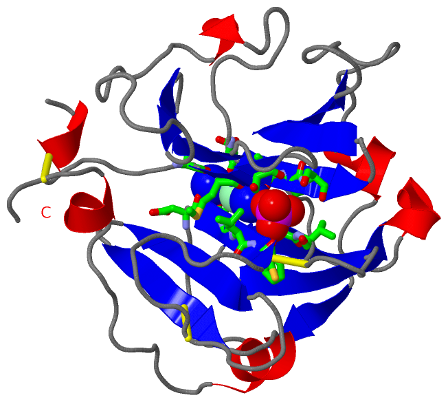
Sites (4, 4)
Asymmetric Unit (4, 4)
|
SS Bonds (3, 3)
Asymmetric/Biological Unit
|
||||||||||||||||
Cis Peptide Bonds (1, 1)
Asymmetric/Biological Unit
|
||||||||
SAPs(SNPs)/Variants (0, 0)| (no "SAP(SNP)/Variant" information available for 1HXN) |
PROSITE Motifs (2, 5)
Asymmetric/Biological Unit (2, 5)
|
||||||||||||||||||||||||||||||||
Exons (0, 0)| (no "Exon" information available for 1HXN) |
Sequences/Alignments
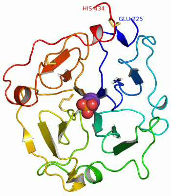
Asymmetric/Biological UnitChain A from PDB Type:PROTEIN Length:210 aligned with HEMO_RABIT | P20058 from UniProtKB/Swiss-Prot Length:460 Alignment length:210 260 270 280 290 300 310 320 330 340 350 360 370 380 390 400 410 420 430 440 450 460 HEMO_RABIT 251 ESTRCDPDLVLSAMVSDNHGATYVFSGSHYWRLDTNRDGWHSWPIAHQWPQGPSTVDAAFSWEDKLYLIQDTKVYVFLTKGGYTLVNGYPKRLEKELGSPPVISLEAVDAAFVCPGSSRLHIMAGRRLWWLDLKSGAQATWTELPWPHEKVDGALCMEKPLGPNSCSTSGPNLYLIHGPNLYCYRHVDKLNAAKNLPQPQRVSRLLGCTH 460 SCOP domains d1hxna_ A: Hemopexin SCOP domains CATH domains 1hxnA00 A:225-434 Hemopexin CATH domains Pfam domains ------------------------------------------------------------------------------------------------------------------------------------------------------------------------------------------------------------------ Pfam domains SAPs(SNPs) ------------------------------------------------------------------------------------------------------------------------------------------------------------------------------------------------------------------ SAPs(SNPs) PROSITE (1) ------HEMOPEXIN_2 PDB: A:231-276 UniProt: 257-302 HEMOPEXIN_2 PDB: A:277-324 UniProt: 303-350 ----HEMOPEXIN_2 PDB: A:329-368 ---HEMOPEXIN_2 PDB: A:372-422 UniProt: 398-448 ------------ PROSITE (1) PROSITE (2) --------------------------------------------HEMOPEXIN ------------------------------------------------------------------------------------------------------------------------------------------------------ PROSITE (2) Transcript ------------------------------------------------------------------------------------------------------------------------------------------------------------------------------------------------------------------ Transcript 1hxn A 225 ESTRCDPDLVLSAMVSDNHGATYVFSGSHYWRLDTNRDGWHSWPIAHQWPQGPSTVDAAFSWEDKLYLIQDTKVYVFLTKGGYTLVNGYPKRLEKELGSPPVISLEAVDAAFVCPGSSRLHIMAGRRLWWLDLKSGAQATWTELPWPHEKVDGALCMEKPLGPNSCSTSGPNLYLIHGPNLYCYRHVDKLNAAKNLPQPQRVSRLLGCTH 434 234 244 254 264 274 284 294 304 314 324 334 344 354 364 374 384 394 404 414 424 434
|
||||||||||||||||||||
SCOP Domains (1, 1)
Asymmetric/Biological Unit
 |
CATH Domains (1, 1)
Asymmetric/Biological Unit
|
Pfam Domains (0, 0)| (no "Pfam Domain" information available for 1HXN) |
Gene Ontology (8, 8)|
Asymmetric/Biological Unit(hide GO term definitions) Chain A (HEMO_RABIT | P20058)
|
||||||||||||||||||||||||||||||||||||||||||||||||||||||||||||||||||
Interactive Views
|
||||||||||||||||||||||||||||||||||||||||||||||||||||||||||||||||||||||||||||||||||||||||||||||||||||||||||||||||||||||||||||||||||||||||||||||||||||||||||
Still Images
|
||||||||||||||||
Databases
|
||||||||||||||||||||||||||||||||||||||||||||||||||||||||||||||||||||||||||||||||||||||||||||||||||||||||||||||||||||||||||||||||||||||||||||||||||||||||||||||||
Analysis Tools
|
|||||||||||||||||||||||||||||||||||||||||||||||||||||||||||||
Entries Sharing at Least One Protein Chain (UniProt ID)
Related Entries Specified in the PDB File
|
|