|
|
|
|
 Description
|
|
Compounds
|
||||||||||||||||||||||||||||||||
Chains, Units
Summary Information (see also Sequences/Alignments below) |
Ligands, Modified Residues, Ions (4, 8)| Asymmetric/Biological Unit (4, 8) |
Sites (9, 9)
Asymmetric Unit (9, 9)
|
SS Bonds (6, 6)
Asymmetric/Biological Unit
|
||||||||||||||||||||||||||||
Cis Peptide Bonds (3, 3)
Asymmetric/Biological Unit
|
||||||||||||||||
SAPs(SNPs)/Variants (0, 0)| (no "SAP(SNP)/Variant" information available for 1QHU) |
PROSITE Motifs (2, 9)
Asymmetric/Biological Unit (2, 9)
|
||||||||||||||||||||||||||||||||
Exons (0, 0)| (no "Exon" information available for 1QHU) |
Sequences/Alignments
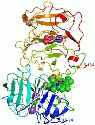
Asymmetric/Biological UnitChain A from PDB Type:PROTEIN Length:398 aligned with HEMO_RABIT | P20058 from UniProtKB/Swiss-Prot Length:460 Alignment length:412 58 68 78 88 98 108 118 128 138 148 158 168 178 188 198 208 218 228 238 248 258 268 278 288 298 308 318 328 338 348 358 368 378 388 398 408 418 428 438 448 458 HEMO_RABIT 49 IEQCSDGWSFDATTLDDNGTMLFFKDEFVWKSHRGIRELISERWKNFIGPVDAAFRHGHTSVYLIKGDKVWVYTSEKNEKVYPKSLQDEFPGIPFPLDAAVECHRGECQDEGILFFQGNRKWFWDLTTGTKKERSWPAVGNCTSALRWLGRYYCFQGNQFLRFNPVSGEVPPGYPLDVRDYFLSCPGRGHRSSHRNSTQHGHESTRCDPDLVLSAMVSDNHGATYVFSGSHYWRLDTNRDGWHSWPIAHQWPQGPSTVDAAFSWEDKLYLIQDTKVYVFLTKGGYTLVNGYPKRLEKELGSPPVISLEAVDAAFVCPGSSRLHIMAGRRLWWLDLKSGAQATWTELPWPHEKVDGALCMEKPLGPNSCSTSGPNLYLIHGPNLYCYRHVDKLNAAKNLPQPQRVSRLLGCTH 460 SCOP domains d1qhua1 A:24-215 Hemopexin d1qhua2 A:222-434 Hemopexin SCOP domains CATH domains 1qhuA01 A:24-213 Hemopexin -- 1qhuA02 A:222-433 Hemopexin - CATH domains Pfam domains (1) -----------------------------------------------------------------------------------------------------------------------------------------------------------------------------------------------------------------------------------------------------------------Hemopexin-1qhuA01 A:280-321 ----------------------------------------------------------------------------------------------------------------- Pfam domains (1) Pfam domains (2) -----------------------------------------------------------------------------------------------------------------------------------------------------------------------------------------------------------------------------------------------------------------Hemopexin-1qhuA02 A:280-321 ----------------------------------------------------------------------------------------------------------------- Pfam domains (2) Pfam domains (3) -----------------------------------------------------------------------------------------------------------------------------------------------------------------------------------------------------------------------------------------------------------------Hemopexin-1qhuA03 A:280-321 ----------------------------------------------------------------------------------------------------------------- Pfam domains (3) Pfam domains (4) -----------------------------------------------------------------------------------------------------------------------------------------------------------------------------------------------------------------------------------------------------------------Hemopexin-1qhuA04 A:280-321 ----------------------------------------------------------------------------------------------------------------- Pfam domains (4) SAPs(SNPs) ---------------------------------------------------------------------------------------------------------------------------------------------------------------------------------------------------------------------------------------------------------------------------------------------------------------------------------------------------------------------------------------------------------------------------- SAPs(SNPs) PROSITE (1) ------HEMOPEXIN_2 PDB: A:30-70 UniProt: 55-95 HEMOPEXIN_2 PDB: A:71-116 UniProt: 96-141 HEMOPEXIN_2 PDB: A:117-161 UniProt: 142-186 HEMOPEXIN_2 PDB: A:162-208 UniProt: 187-233 -----------------------HEMOPEXIN_2 PDB: A:231-276 UniProt: 257-302 HEMOPEXIN_2 PDB: A:277-324 UniProt: 303-350 ----HEMOPEXIN_2 PDB: A:329-368 ---HEMOPEXIN_2 PDB: A:372-422 UniProt: 398-448 ------------ PROSITE (1) PROSITE (2) ------------------------------------------------------------------------------------------------------------------------------------------------------------------------------------------------------------------------------------------------------HEMOPEXIN ------------------------------------------------------------------------------------------------------------------------------------------------------ PROSITE (2) Transcript ---------------------------------------------------------------------------------------------------------------------------------------------------------------------------------------------------------------------------------------------------------------------------------------------------------------------------------------------------------------------------------------------------------------------------- Transcript 1qhu A 24 IEQCSDGWSFDATTLDDNGTMLFFKDEFVWKSHRGIRELISERWKNFIGPVDAAFRHGHTSVYLIKGDKVWVYTS-------PKSLQDEFPGIPFPLDAAVECHRGECQDEGILFFQGNRKWFWDLTTGTKKERSWPAVGNCTSALRWLGRYYCFQGNQFLRFNPVSGEVPPGYPLDVRDYFLSCPGRGHRS-------HGHESTRCDPDLVLSAMVSDNHGATYVFSGSHYWRLDTNRDGWHSWPIAHQWPQGPSTVDAAFSWEDKLYLIQDTKVYVFLTKGGYTLVNGYPKRLEKELGSPPVISLEAVDAAFVCPGSSRLHIMAGRRLWWLDLKSGAQATWTELPWPHEKVDGALCMEKPLGPNSCSTSGPNLYLIHGPNLYCYRHVDKLNAAKNLPQPQRVSRLLGCTH 434 33 43 53 63 73 83 93 | - | 113 123 133 143 153 163 173 183 193 203 213 | 222 232 242 252 262 272 282 292 302 312 322 332 342 352 362 372 382 392 402 412 422 432 98 106 215 222
|
||||||||||||||||||||
SCOP Domains (1, 2)
Asymmetric/Biological Unit
 |
CATH Domains (1, 2)
Asymmetric/Biological Unit
|
Pfam Domains (1, 4)
Asymmetric/Biological Unit
|
Gene Ontology (8, 8)|
Asymmetric/Biological Unit(hide GO term definitions) Chain A (HEMO_RABIT | P20058)
|
||||||||||||||||||||||||||||||||||||||||||||||||||||||||||||||||||
Interactive Views
|
||||||||||||||||||||||||||||||||||||||||||||||||||||||||||||||||||||||||||||||||||||||||||||||||||||||||||||||||||||||||||||||||||||||||||||||||||||||||||||||||||||||||||||||||||||||||||||||||||||||||||||||||||
Still Images
|
||||||||||||||||
Databases
|
||||||||||||||||||||||||||||||||||||||||||||||||||||||||||||||||||||||||||||||||||||||||||||||||||||||||||||||||||||||||||||||||||||||||||||||||||||||||||||||||
Analysis Tools
|
|||||||||||||||||||||||||||||||||||||||||||||||||||||||||||||
Entries Sharing at Least One Protein Chain (UniProt ID)
Related Entries Specified in the PDB File
|
|