Show PDB file:   
         Plain Text   HTML   (compressed file size)
QuickSearch:   
by PDB,NDB,UniProt,PROSITE Code or Search Term(s)  
(-)Asymmetric Unit
(-)Asym. Unit - sites
(-)Biological Unit 1
(-)Biol. Unit 1 - sites
collapse expand < >
Image Asymmetric Unit
Asymmetric Unit  (Jmol Viewer)
Image Asym. Unit - sites
Asym. Unit - sites  (Jmol Viewer)
Image Biological Unit 1
Biological Unit 1  (Jmol Viewer)
Image Biol. Unit 1 - sites
Biol. Unit 1 - sites  (Jmol Viewer)

(-) Description

Title :  CARBOHYDRATE BINDING MODULE FAMILY29 COMPLEXED WITH MANNOHEXAOSE
 
Authors :  S. J. Charnock, D. Nurizzo, G. J. Davies
Date :  19 Mar 02  (Deposition) - 20 Mar 03  (Release) - 13 Jul 11  (Revision)
Method :  X-RAY DIFFRACTION
Resolution :  1.51
Chains :  Asym. Unit :  A
Biol. Unit 1:  A  (2x)
Keywords :  Carbohydrate Binding Domain, Glucomannan, Cellohexaose, Mannohexaose, Cellulosome (Keyword Search: [Gene Ontology, PubMed, Web (Google))
 
Reference :  S. J. Charnock, D. Bolam, D. Nurizzo, L. Szabo, V. Mckie, H. Gilbert, G. J. Davies
Promiscuity In Ligand-Binding: The Three-Dimensional Structure Of A Piromyces Carbohydrate-Binding Module, Cbm29-2, In Complex With Cello- And Mannohexaose
Proc. Natl. Acad. Sci. Usa V. 99 14077 2002
PubMed-ID: 12391332  |  Reference-DOI: 10.1073/PNAS.212516199

(-) Compounds

Molecule 1 - NON-CATALYTIC PROTEIN 1
    ChainsA
    EngineeredYES
    Expression SystemESCHERICHIA COLI
    Expression System PlasmidPET22B
    Expression System StrainBL21
    Expression System Taxid511693
    FragmentCARBOHYDRATE BINDING MODULE FAMILY 29 RESIDUES 335-478
    Organism ScientificPIROMYCES EQUI
    Organism Taxid99929
    SynonymNCP1

 Structural Features

(-) Chains, Units

  1
Asymmetric Unit A
Biological Unit 1 (2x)A

Summary Information (see also Sequences/Alignments below)

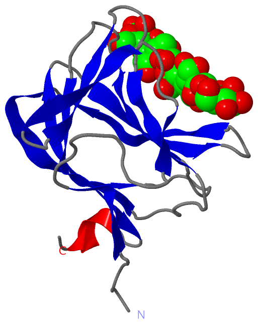
(-) Ligands, Modified Residues, Ions  (1, 6)

Asymmetric Unit (1, 6)
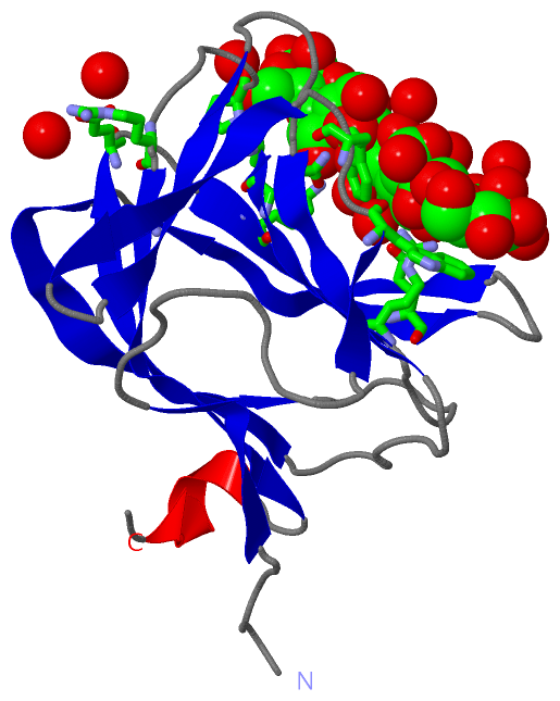
No.NameCountTypeFull Name
1BMA6Ligand/IonBETA-D-MANNOSE
Biological Unit 1 (1, 12)
No.NameCountTypeFull Name
1BMA12Ligand/IonBETA-D-MANNOSE

(-) Sites  (6, 6)

Asymmetric Unit (6, 6)
No.NameEvidenceResiduesDescription
1AC1SOFTWAREALA A:118 , PRO A:119 , ARG A:124 , BMA A:1144 , HOH A:2109 , HOH A:2189BINDING SITE FOR RESIDUE BMA A1143
2AC2SOFTWAREGLU A:70 , LYS A:74 , GLN A:116 , ALA A:118 , BMA A:1143 , BMA A:1145 , HOH A:2169BINDING SITE FOR RESIDUE BMA A1144
3AC3SOFTWARETRP A:26 , GLN A:116 , BMA A:1144 , BMA A:1146 , HOH A:2191 , HOH A:2192 , HOH A:2193 , HOH A:2194BINDING SITE FOR RESIDUE BMA A1145
4AC4SOFTWARETRP A:26 , GLU A:78 , ARG A:112 , BMA A:1145 , BMA A:1147 , HOH A:2119 , HOH A:2192 , HOH A:2195 , HOH A:2196BINDING SITE FOR RESIDUE BMA A1146
5AC5SOFTWARETRP A:24 , ARG A:112 , BMA A:1146 , BMA A:1148 , HOH A:2119 , HOH A:2198 , HOH A:2199BINDING SITE FOR RESIDUE BMA A1147
6AC6SOFTWARETRP A:24 , SER A:92 , BMA A:1147 , HOH A:2200BINDING SITE FOR RESIDUE BMA A1148

(-) SS Bonds  (0, 0)

(no "SS Bond" information available for 1GWL)

(-) Cis Peptide Bonds  (0, 0)

(no "Cis Peptide Bond" information available for 1GWL)

 Sequence-Structure Mapping

(-) SAPs(SNPs)/Variants  (0, 0)

(no "SAP(SNP)/Variant" information available for 1GWL)

(-) PROSITE Motifs  (0, 0)

(no "PROSITE Motif" information available for 1GWL)

(-) Exons   (0, 0)

(no "Exon" information available for 1GWL)

(-) Sequences/Alignments

Asymmetric Unit
   Reformat: Number of residues per line =  ('0' or empty: single-line sequence representation)
  Number of residues per labelling interval =   
  UniProt sequence: complete  aligned part    
   Show mapping: SCOP domains CATH domains Pfam domains Secondary structure (by author)
SAPs(SNPs) PROSITE motifs Exons
(details for a mapped element are shown in a popup box when the mouse pointer rests over it)
Chain A from PDB  Type:PROTEIN  Length:141
 aligned with Q9C171_PIREQ | Q9C171 from UniProtKB/TrEMBL  Length:478

    Alignment length:141
                                   344       354       364       374       384       394       404       414       424       434       444       454       464       474 
         Q9C171_PIREQ   335 NVRATYTVIFKNASGLPNGYDNWGWGCTLSYYGGAMIINPQEGKYGAVSLKRNSGSFRGGSLRFDMKNEGKVKILVENSEADEKFEVETISPSDEYVTYILDVDFDLPFDRIDFQDAPGNGDRIWIKNLVHSTGSADDFVD 475
               SCOP domains d1gwla_ A: Non-catalytic protein 1, Ncp1                                                                                                      SCOP domains
               CATH domains 1gwlA00 A:2-142  [code=2.60.120.360, no name defined]                                                                                         CATH domains
               Pfam domains --------------------------------------------------------------------------------------------------------------------------------------------- Pfam domains
         Sec.struct. author .....eeeee.........eee....eeeeee..eeeeee......eeeeee........eeeeeeee...eeeeeee....eeeeeeee..eeeeeeeeee........eeeeee.......eeeeeeeee..hhhhh.. Sec.struct. author
                 SAPs(SNPs) --------------------------------------------------------------------------------------------------------------------------------------------- SAPs(SNPs)
                    PROSITE --------------------------------------------------------------------------------------------------------------------------------------------- PROSITE
                 Transcript --------------------------------------------------------------------------------------------------------------------------------------------- Transcript
                 1gwl A   2 NVRATYTVIFKNASGLPNGYDNWGWGCTLSYYGGAMIINPQEGKYGAVSLKRNSGSFRGGSLRFDMKNEGKVKILVENSEADEKFEVETISPSDEYVTYILDVDFDLPFDRIDFQDAPGNGDRIWIKNLVHSTGSADDFVD 142
                                    11        21        31        41        51        61        71        81        91       101       111       121       131       141 

   Legend:   → Mismatch (orange background)
  - → Gap (green background, '-', border residues have a numbering label)
    → Modified Residue (blue background, lower-case, 'x' indicates undefined single-letter code, labelled with number + name)
  x → Chemical Group (purple background, 'x', labelled with number + name, e.g. ACE or NH2)
  extra numbering lines below/above indicate numbering irregularities and modified residue names etc., number ends below/above '|'

 Classification and Annotation

(-) SCOP Domains  (1, 1)

Asymmetric Unit

(-) CATH Domains  (1, 1)

Asymmetric Unit
(-)
Class: Mainly Beta (13760)

(-) Pfam Domains  (0, 0)

(no "Pfam Domain" information available for 1GWL)

(-) Gene Ontology  (0, 0)

Asymmetric Unit(hide GO term definitions)
    (no "Gene Ontology" information available for 1GWL)

 Visualization

(-) Interactive Views

Asymmetric Unit
  Complete Structure
    Jena3D(integrated viewing of ligand, site, SAP, PROSITE, SCOP information)
    WebMol | AstexViewer[tm]@PDBe
(Java Applets, require no local installation except for Java; loading may be slow)
    STRAP
(Java WebStart application, automatic local installation, requires Java; full application with system access!)
    RasMol
(require local installation)
    Molscript (VRML)
(requires installation of a VRML viewer; select preferred view via VRML and generate a mono or stereo PDF format file)
 
  Ligands, Modified Residues, Ions
    BMA  [ RasMol | Jena3D ]  +environment [ RasMol | Jena3D ]
 
  Sites
    AC1  [ RasMol ]  +environment [ RasMol ]
    AC2  [ RasMol ]  +environment [ RasMol ]
    AC3  [ RasMol ]  +environment [ RasMol ]
    AC4  [ RasMol ]  +environment [ RasMol ]
    AC5  [ RasMol ]  +environment [ RasMol ]
    AC6  [ RasMol ]  +environment [ RasMol ]
 
  Cis Peptide Bonds
(no "Cis Peptide Bonds" information available for 1gwl)
 
Biological Unit
  Complete Structure
    Biological Unit 1  [ Jena3D ]

(-) Still Images

Jmol
  protein: cartoon or spacefill or dots and stick; nucleic acid: cartoon and stick; ligands: spacefill; active site: stick
Molscript
  protein, nucleic acid: cartoon; ligands: spacefill; active site: ball and stick

 Databases and Analysis Tools

(-) Databases

Access by PDB/NDB ID
  1gwl
    Family and Domain InformationProDom | SYSTERS
    General Structural InformationGlycoscienceDB | MMDB | NDB | OCA | PDB | PDBe | PDBj | PDBsum | PDBWiki | PQS | PROTEOPEDIA
    Orientation in MembranesOPM
    Protein SurfaceSURFACE
    Secondary StructureDSSP (structure derived) | HSSP (homology derived)
    Structural GenomicsGeneCensus
    Structural NeighboursCE | VAST
    Structure ClassificationCATH | Dali | SCOP
    Validation and Original DataBMRB Data View | BMRB Restraints Grid | EDS | PROCHECK | RECOORD | WHAT_CHECK
 
Access by UniProt ID/Accession number
  Q9C171_PIREQ | Q9C171
    Comparative Protein Structure ModelsModBase
    Genomic InformationEnsembl
    Protein-protein InteractionDIP
    Sequence, Family and Domain InformationInterPro | Pfam | SMART | UniProtKB/TrEMBL
 
Access by Enzyme Classificator   (EC Number)
  (no 'Enzyme Classificator' available)
    General Enzyme InformationBRENDA | EC-PDB | Enzyme | IntEnz
    PathwayKEGG | MetaCyc
 
Access by Disease Identifier   (MIM ID)
  (no 'MIM ID' available)
    Disease InformationOMIM
 
Access by GenAge ID
  (no 'GenAge ID' available)
    Age Related InformationGenAge

(-) Analysis Tools

Access by PDB/NDB ID
    Domain InformationXDom
    Interatomic Contacts of Structural UnitsCSU
    Ligand-protein ContactsLPC
    Protein CavitiescastP
    Sequence and Secondary StructurePDBCartoon
    Structure AlignmentSTRAP(Java WebStart application, automatic local installation, requires Java; full application with system access!)
    Structure and Sequence BrowserSTING
 
Access by UniProt ID/Accession number
  Q9C171_PIREQ | Q9C171
    Protein Disorder PredictionDisEMBL | FoldIndex | GLOBPLOT (for more information see DisProt)

 Related Entries

(-) Entries Sharing at Least One Protein Chain (UniProt ID)

UniProtKB/TrEMBL
        Q9C171_PIREQ | Q9C1711gwk 1gwm 1oh3 1w8t 1w8u 1w8w 1w8z 1w90 1w9f 1wcu

(-) Related Entries Specified in the PDB File

1gwk CARBOHYDRATE BINDING MODULE FAMILY29
1gwm CARBOHYDRATE BINDING MODULE FAMILY29 COMPLEXED WITH GLUCOHEXAOSE