Show PDB file:   
         Plain Text   HTML   (compressed file size)
QuickSearch:   
by PDB,NDB,UniProt,PROSITE Code or Search Term(s)  
(-)Asymmetric Unit
(-)Asym. Unit - sites
(-)Biological Unit 1
(-)Biol. Unit 1 - sites
(-)Biological Unit 2
collapse expand < >
Image Asymmetric Unit
Asymmetric Unit  (Jmol Viewer)
Image Asym. Unit - sites
Asym. Unit - sites  (Jmol Viewer)
Image Biological Unit 1
Biological Unit 1  (Jmol Viewer)
Image Biol. Unit 1 - sites
Biol. Unit 1 - sites  (Jmol Viewer)
Image Biological Unit 2
Biological Unit 2  (Jmol Viewer)

(-) Description

Title :  RIBONUCLEASE BI(G SPECIFIC ENDONUCLEASE) COMPLEXED WITH SULFATE IONS
 
Authors :  K. M. Polyakov, A. A. Lebedev, A. G. Pavlovsky, R. G. Sanishvili, G. G. Dodson
Date :  26 Oct 01  (Deposition) - 29 Nov 01  (Release) - 24 Feb 09  (Revision)
Method :  X-RAY DIFFRACTION
Resolution :  2.00
Chains :  Asym. Unit :  A,B
Biol. Unit 1:  A  (1x)
Biol. Unit 2:  B  (1x)
Keywords :  Hydrolase, Endoribonuclease, Nuclease (Keyword Search: [Gene Ontology, PubMed, Web (Google))
 
Reference :  K. M. Polyakov, A. A. Lebedev, A. L. Okorokov, K. I. Panov, A. A. Schulga, A. G. Pavlovsky, M. Y. A. Karpeisky, G. G. Dodson
The Structure Of Substrate-Free Microbial Ribonuclease Binase And Of Its Complexes With 3'Gmp And Sulfate Ions
Acta Crystallogr. , Sect. D V. 58 744 2002
PubMed-ID: 11976484  |  Reference-DOI: 10.1107/S0907444902003207
(for further references see the PDB file header)

(-) Compounds

Molecule 1 - RIBONUCLEASE
    ChainsA, B
    EC Number3.1.27.3
    Organism ScientificBACILLUS INTERMEDIUS
    Organism Taxid1400
    Other DetailsRIBONUCLEASE BI(G SPECIFIC ENDONUCLEASE) COMPLEXED WITH SULFATE IONS
    SynonymBINASE

 Structural Features

(-) Chains, Units

  12
Asymmetric Unit AB
Biological Unit 1 (1x)A 
Biological Unit 2 (1x) B

Summary Information (see also Sequences/Alignments below)

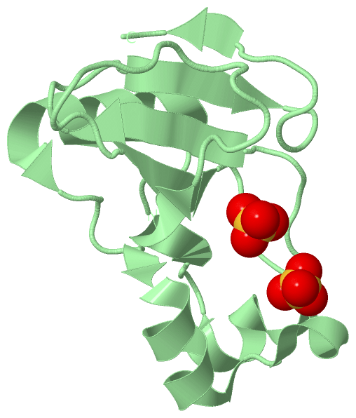
(-) Ligands, Modified Residues, Ions  (1, 4)

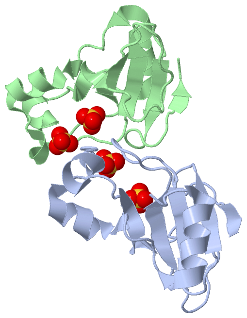
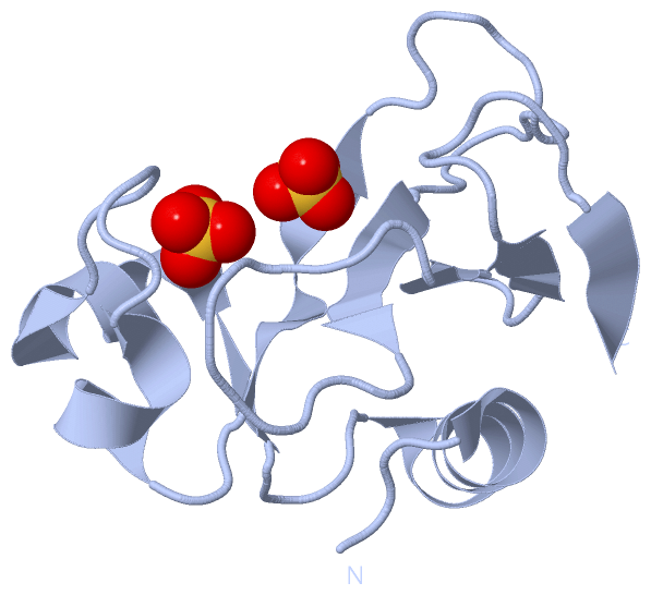
Asymmetric Unit (1, 4)
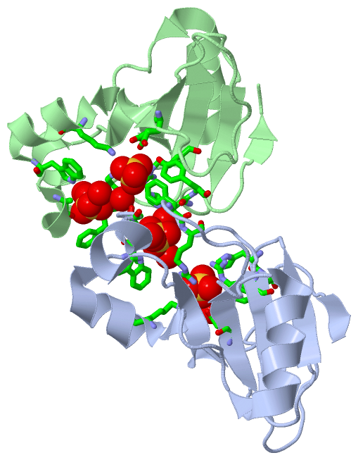
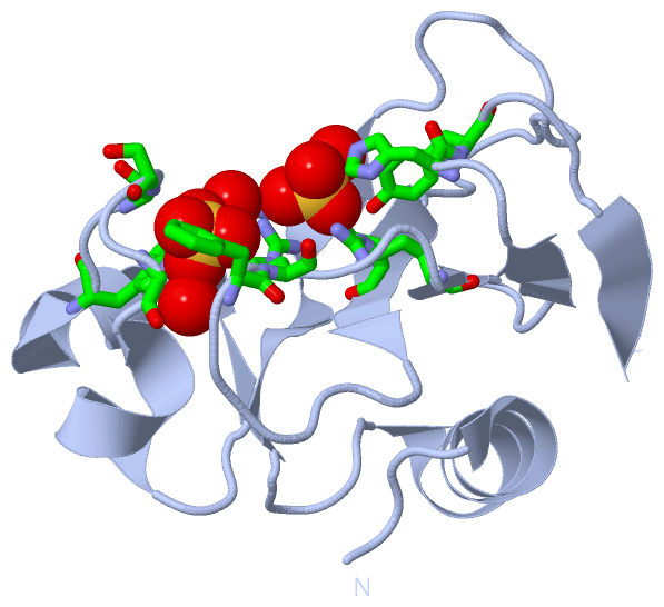
No.NameCountTypeFull Name
1SO44Ligand/IonSULFATE ION
Biological Unit 1 (1, 2)
No.NameCountTypeFull Name
1SO42Ligand/IonSULFATE ION
Biological Unit 2 (1, 2)
No.NameCountTypeFull Name
1SO42Ligand/IonSULFATE ION

(-) Sites  (4, 4)

Asymmetric Unit (4, 4)
No.NameEvidenceResiduesDescription
1AC1SOFTWARELYS A:26 , GLU A:72 , ARG A:82 , ARG A:86 , HIS A:101 , TYR A:102 , HOH A:2086BINDING SITE FOR RESIDUE SO4 A1110
2AC2SOFTWARETRP A:34 , ALA A:36 , SER A:37 , GLY A:39 , PHE A:81 , ARG A:82 , HOH A:2087 , HOH A:2088 , HOH B:2085BINDING SITE FOR RESIDUE SO4 A1111
3AC3SOFTWARELYS B:26 , GLU B:72 , ARG B:82 , ARG B:86 , HIS B:101 , TYR B:102 , HOH B:2092BINDING SITE FOR RESIDUE SO4 B1110
4AC4SOFTWARETRP B:34 , ALA B:36 , GLY B:39 , PHE B:81 , ARG B:82 , HOH B:2093 , HOH B:2094BINDING SITE FOR RESIDUE SO4 B1111

(-) SS Bonds  (0, 0)

(no "SS Bond" information available for 1GOV)

(-) Cis Peptide Bonds  (0, 0)

(no "Cis Peptide Bond" information available for 1GOV)

 Sequence-Structure Mapping

(-) SAPs(SNPs)/Variants  (0, 0)

(no "SAP(SNP)/Variant" information available for 1GOV)

(-) PROSITE Motifs  (0, 0)

(no "PROSITE Motif" information available for 1GOV)

(-) Exons   (0, 0)

(no "Exon" information available for 1GOV)

(-) Sequences/Alignments

Asymmetric Unit
   Reformat: Number of residues per line =  ('0' or empty: single-line sequence representation)
  Number of residues per labelling interval =   
  UniProt sequence: complete  aligned part    
   Show mapping: SCOP domains CATH domains Pfam domains Secondary structure (by author)
SAPs(SNPs) PROSITE motifs Exons
(details for a mapped element are shown in a popup box when the mouse pointer rests over it)
Chain A from PDB  Type:PROTEIN  Length:108
 aligned with RN_BACIN | P00649 from UniProtKB/Swiss-Prot  Length:162

    Alignment length:108
                                    64        74        84        94       104       114       124       134       144       154        
             RN_BACIN    55 VINTFDGVADYLIRYKRLPDNYITKSQASALGWVASKGNLAEVAPGKSIGGDVFSNREGRLPSASGRTWREADINYVSGFRNADRLVYSSDWLIYKTTDHYATFTRIR 162
               SCOP domains d1gova_ A: Binase                                                                                            SCOP domains
               CATH domains 1govA00 A:2-109  [code=3.10.450.30, no name defined]                                                         CATH domains
               Pfam domains ------------------------------------------------------------------------------------------------------------ Pfam domains
         Sec.struct. author ...hhhhhhhhhhhhh.....eehhhhhhhhh......hhhhhh...eeeeeee..............eeeee...........eeeee....eeee.......eeee Sec.struct. author
                 SAPs(SNPs) ------------------------------------------------------------------------------------------------------------ SAPs(SNPs)
                    PROSITE ------------------------------------------------------------------------------------------------------------ PROSITE
                 Transcript ------------------------------------------------------------------------------------------------------------ Transcript
                 1gov A   2 VINTFDGVADYLIRYKRLPNDYITKSQASALGWVASKGDLAEVAPGKSIGGDVFSNREGRLPSAGSRTWREADINYVSGFRNADRLVYSSDWLIYKTTDHYATFTRIR 109
                                    11        21        31        41        51        61        71        81        91       101        

Chain B from PDB  Type:PROTEIN  Length:108
 aligned with RN_BACIN | P00649 from UniProtKB/Swiss-Prot  Length:162

    Alignment length:108
                                    64        74        84        94       104       114       124       134       144       154        
             RN_BACIN    55 VINTFDGVADYLIRYKRLPDNYITKSQASALGWVASKGNLAEVAPGKSIGGDVFSNREGRLPSASGRTWREADINYVSGFRNADRLVYSSDWLIYKTTDHYATFTRIR 162
               SCOP domains d1govb_ B: Binase                                                                                            SCOP domains
               CATH domains 1govB00 B:2-109  [code=3.10.450.30, no name defined]                                                         CATH domains
               Pfam domains ------------------------------------------------------------------------------------------------------------ Pfam domains
         Sec.struct. author ...hhhhhhhhhhhhh.....eehhhhhhhhh.hhhhhhhhhhh...eeeeeee..............eeeee...........eeeee....eeee.......ee.. Sec.struct. author
                 SAPs(SNPs) ------------------------------------------------------------------------------------------------------------ SAPs(SNPs)
                    PROSITE ------------------------------------------------------------------------------------------------------------ PROSITE
                 Transcript ------------------------------------------------------------------------------------------------------------ Transcript
                 1gov B   2 VINTFDGVADYLIRYKRLPNDYITKSQASALGWVASKGDLAEVAPGKSIGGDVFSNREGRLPSAGSRTWREADINYVSGFRNADRLVYSSDWLIYKTTDHYATFTRIR 109
                                    11        21        31        41        51        61        71        81        91       101        

   Legend:   → Mismatch (orange background)
  - → Gap (green background, '-', border residues have a numbering label)
    → Modified Residue (blue background, lower-case, 'x' indicates undefined single-letter code, labelled with number + name)
  x → Chemical Group (purple background, 'x', labelled with number + name, e.g. ACE or NH2)
  extra numbering lines below/above indicate numbering irregularities and modified residue names etc., number ends below/above '|'

 Classification and Annotation

(-) SCOP Domains  (1, 2)

Asymmetric Unit

(-) CATH Domains  (1, 2)

Asymmetric Unit
(-)
Class: Alpha Beta (26913)

(-) Pfam Domains  (0, 0)

(no "Pfam Domain" information available for 1GOV)

(-) Gene Ontology  (10, 10)

Asymmetric Unit(hide GO term definitions)
Chain A,B   (RN_BACIN | P00649)
molecular function
    GO:0003723    RNA binding    Interacting selectively and non-covalently with an RNA molecule or a portion thereof.
    GO:0004519    endonuclease activity    Catalysis of the hydrolysis of ester linkages within nucleic acids by creating internal breaks.
    GO:0004521    endoribonuclease activity    Catalysis of the hydrolysis of ester linkages within ribonucleic acid by creating internal breaks.
    GO:0016787    hydrolase activity    Catalysis of the hydrolysis of various bonds, e.g. C-O, C-N, C-C, phosphoric anhydride bonds, etc. Hydrolase is the systematic name for any enzyme of EC class 3.
    GO:0004518    nuclease activity    Catalysis of the hydrolysis of ester linkages within nucleic acids.
    GO:0004540    ribonuclease activity    Catalysis of the hydrolysis of phosphodiester bonds in chains of RNA.
biological process
    GO:0090501    RNA phosphodiester bond hydrolysis    The RNA metabolic process in which the phosphodiester bonds between ribonucleotides are cleaved by hydrolysis.
    GO:0090502    RNA phosphodiester bond hydrolysis, endonucleolytic    The chemical reactions and pathways involving the hydrolysis of internal 3',5'-phosphodiester bonds in one or two strands of ribonucleotides.
    GO:0090305    nucleic acid phosphodiester bond hydrolysis    The nucleic acid metabolic process in which the phosphodiester bonds between nucleotides are cleaved by hydrolysis.
cellular component
    GO:0005576    extracellular region    The space external to the outermost structure of a cell. For cells without external protective or external encapsulating structures this refers to space outside of the plasma membrane. This term covers the host cell environment outside an intracellular parasite.

 Visualization

(-) Interactive Views

Asymmetric Unit
  Complete Structure
    Jena3D(integrated viewing of ligand, site, SAP, PROSITE, SCOP information)
    WebMol | AstexViewer[tm]@PDBe
(Java Applets, require no local installation except for Java; loading may be slow)
    STRAP
(Java WebStart application, automatic local installation, requires Java; full application with system access!)
    RasMol
(require local installation)
    Molscript (VRML)
(requires installation of a VRML viewer; select preferred view via VRML and generate a mono or stereo PDF format file)
 
  Ligands, Modified Residues, Ions
    SO4  [ RasMol | Jena3D ]  +environment [ RasMol | Jena3D ]
 
  Sites
    AC1  [ RasMol ]  +environment [ RasMol ]
    AC2  [ RasMol ]  +environment [ RasMol ]
    AC3  [ RasMol ]  +environment [ RasMol ]
    AC4  [ RasMol ]  +environment [ RasMol ]
 
  Cis Peptide Bonds
(no "Cis Peptide Bonds" information available for 1gov)
 
Biological Units
  Complete Structure
    Biological Unit 1  [ Jena3D ]
    Biological Unit 2  [ Jena3D ]

(-) Still Images

Jmol
  protein: cartoon or spacefill or dots and stick; nucleic acid: cartoon and stick; ligands: spacefill; active site: stick
Molscript
  protein, nucleic acid: cartoon; ligands: spacefill; active site: ball and stick

 Databases and Analysis Tools

(-) Databases

Access by PDB/NDB ID
  1gov
    Family and Domain InformationProDom | SYSTERS
    General Structural InformationGlycoscienceDB | MMDB | NDB | OCA | PDB | PDBe | PDBj | PDBsum | PDBWiki | PQS | PROTEOPEDIA
    Orientation in MembranesOPM
    Protein SurfaceSURFACE
    Secondary StructureDSSP (structure derived) | HSSP (homology derived)
    Structural GenomicsGeneCensus
    Structural NeighboursCE | VAST
    Structure ClassificationCATH | Dali | SCOP
    Validation and Original DataBMRB Data View | BMRB Restraints Grid | EDS | PROCHECK | RECOORD | WHAT_CHECK
 
Access by UniProt ID/Accession number
  RN_BACIN | P00649
    Comparative Protein Structure ModelsModBase
    Genomic InformationEnsembl
    Protein-protein InteractionDIP
    Sequence, Family and Domain InformationInterPro | Pfam | SMART | UniProtKB/SwissProt
 
Access by Enzyme Classificator   (EC Number)
  3.1.27.3
    General Enzyme InformationBRENDA | EC-PDB | Enzyme | IntEnz
    PathwayKEGG | MetaCyc
 
Access by Disease Identifier   (MIM ID)
  (no 'MIM ID' available)
    Disease InformationOMIM
 
Access by GenAge ID
  (no 'GenAge ID' available)
    Age Related InformationGenAge

(-) Analysis Tools

Access by PDB/NDB ID
    Domain InformationXDom
    Interatomic Contacts of Structural UnitsCSU
    Ligand-protein ContactsLPC
    Protein CavitiescastP
    Sequence and Secondary StructurePDBCartoon
    Structure AlignmentSTRAP(Java WebStart application, automatic local installation, requires Java; full application with system access!)
    Structure and Sequence BrowserSTING
 
Access by UniProt ID/Accession number
  RN_BACIN | P00649
    Protein Disorder PredictionDisEMBL | FoldIndex | GLOBPLOT (for more information see DisProt)

 Related Entries

(-) Entries Sharing at Least One Protein Chain (UniProt ID)

UniProtKB/Swiss-Prot
        RN_BACIN | P006491buj 1gou 1goy 2rbi 4haa

(-) Related Entries Specified in the PDB File

1buj STRUCTURE OF BINASE IN SOLUTION
1gou STRUCTURE OF RIBONUCLEASE BI(G SPECIFIC ENDONUCLEASE) UNLIGANDED FORM
1goy STRUCTURE OF RIBONUCLEASE BI(G SPECIFIC ENDONUCLEASE) COMPLEXED WITH GUANOSINE-3'-PHOSPHATE (3'-GMP)
2rbi STRUCTURE OF BINASE MUTANT HIS 101 ASN