|
|
|
|
 Description
|
|
Compounds
|
||||||||||||||||||||||||
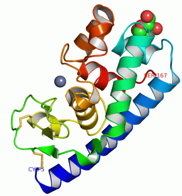
Chains, Units
Summary Information (see also Sequences/Alignments below) |
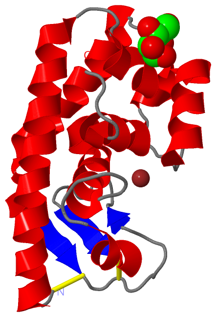
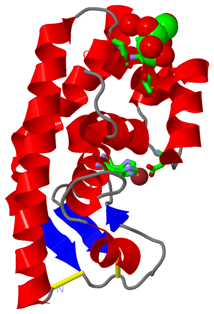
Ligands, Modified Residues, Ions (2, 2)| Asymmetric/Biological Unit (2, 2) |
Sites (2, 2)
Asymmetric Unit (2, 2)
|
SS Bonds (2, 2)
Asymmetric/Biological Unit
|
||||||||||||
Cis Peptide Bonds (1, 1)
Asymmetric/Biological Unit
|
||||||||
SAPs(SNPs)/Variants (0, 0)| (no "SAP(SNP)/Variant" information available for 1GE6) |
PROSITE Motifs (0, 0)| (no "PROSITE Motif" information available for 1GE6) |
Exons (0, 0)| (no "Exon" information available for 1GE6) |
Sequences/Alignments
Asymmetric/Biological UnitChain A from PDB Type:PROTEIN Length:163 aligned with PLMP_GRIFR | P81054 from UniProtKB/Swiss-Prot Length:348 Alignment length:163 195 205 215 225 235 245 255 265 275 285 295 305 315 325 335 345 PLMP_GRIFR 186 CSSSEQSALAAAASAAQSYVAESLSYLQTHTAATPRYTTWFGSYISSRHSTVLQHYTDMNSNDFSSYSFDCTCTAAGTFAYVYPNRFGTVYLCGAFWKAPTTGTDSQAGTLVHESSHFTRNGGTKDYAYGQAAAKSLATMDPDKAVMNADNHEYFSENNPAQS 348 SCOP domains d1ge6a_ A: Fungal zinc peptidase SCOP domains CATH domains 1ge6A00 A:5-167 Collagenase (Catalytic Domain) CATH domains Pfam domains ------------------------------------------------------------------------------------------------------------------------------------------------------------------- Pfam domains SAPs(SNPs) ------------------------------------------------------------------------------------------------------------------------------------------------------------------- SAPs(SNPs) PROSITE ------------------------------------------------------------------------------------------------------------------------------------------------------------------- PROSITE Transcript ------------------------------------------------------------------------------------------------------------------------------------------------------------------- Transcript 1ge6 A 5 CSSSEQSALAAAASAAQSYVAESLSYLQTHTAATPRYTTWFGSYISSRHSTVLQHYTDMNSNDFSSYSFDCTCTAAGTFAYVYPNRFGTVYLCGAFWKAPTTGTDSQAGTLVHESSHFTRNGGTKDYAYGQAAAKSLATMDPDKAVMNADNHEYFSENNPAQS 167 14 24 34 44 54 64 74 84 94 104 114 124 134 144 154 164
|
||||||||||||||||||||
SCOP Domains (1, 1)
Asymmetric/Biological Unit
 |
CATH Domains (1, 1)
Asymmetric/Biological Unit
|
Pfam Domains (0, 0)| (no "Pfam Domain" information available for 1GE6) |
Gene Ontology (7, 7)|
Asymmetric/Biological Unit(hide GO term definitions) Chain A (PLMP_GRIFR | P81054)
|
||||||||||||||||||||||||||||||||||||||||||||||||||||||||||||
Interactive Views
|
|||||||||||||||||||||||||||||||||||||||||||||||||||||||||||||||||||||||||||||||||||||||||||||||||||||||||||||||||||||||||||||||||||||
Still Images
|
||||||||||||||||
Databases
|
||||||||||||||||||||||||||||||||||||||||||||||||||||||||||||||||||||||||||||||||||||||||||||||||||||||||||||||||||||||||||||||||||||||||||||||||||||||||||||||||
Analysis Tools
|
|||||||||||||||||||||||||||||||||||||||||||||||||||||||||||||
Entries Sharing at Least One Protein Chain (UniProt ID)
Related Entries Specified in the PDB File
|
|