| molecular function |
|---|
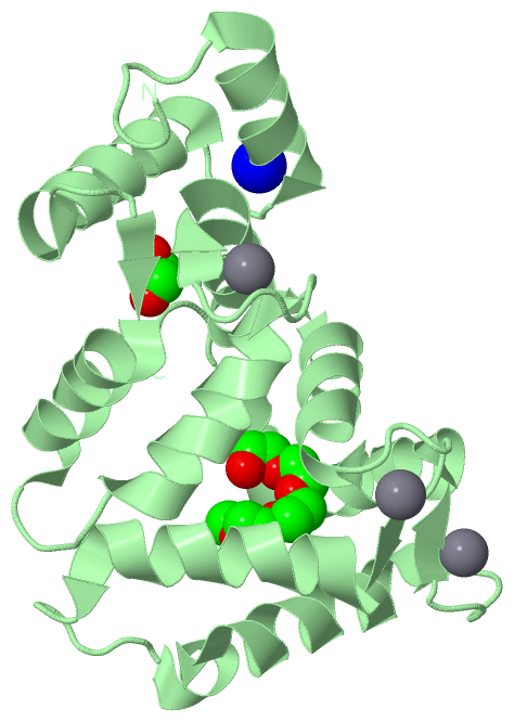
| | GO:0005509 | | calcium ion binding | | Interacting selectively and non-covalently with calcium ions (Ca2+). |
| | GO:0000287 | | magnesium ion binding | | Interacting selectively and non-covalently with magnesium (Mg) ions. |
| | GO:0046872 | | metal ion binding | | Interacting selectively and non-covalently with any metal ion. |
| | GO:0005515 | | protein binding | | Interacting selectively and non-covalently with any protein or protein complex (a complex of two or more proteins that may include other nonprotein molecules). |
| | GO:0019901 | | protein kinase binding | | Interacting selectively and non-covalently with a protein kinase, any enzyme that catalyzes the transfer of a phosphate group, usually from ATP, to a protein substrate. |
| | GO:0005245 | | voltage-gated calcium channel activity | | Enables the transmembrane transfer of a calcium ion by a voltage-gated channel. A voltage-gated channel is a channel whose open state is dependent on the voltage across the membrane in which it is embedded. |
| biological process |
|---|
| | GO:0070588 | | calcium ion transmembrane transport | | A process in which a calcium ion is transported from one side of a membrane to the other by means of some agent such as a transporter or pore. |
| | GO:0048015 | | phosphatidylinositol-mediated signaling | | A series of molecular signals in which a cell uses a phosphatidylinositol-mediated signaling to convert a signal into a response. Phosphatidylinositols include phosphatidylinositol (PtdIns) and its phosphorylated derivatives. |
| | GO:0045921 | | positive regulation of exocytosis | | Any process that activates or increases the frequency, rate or extent of exocytosis. |
| | GO:0010975 | | regulation of neuron projection development | | Any process that modulates the rate, frequency or extent of neuron projection development. Neuron projection development is the process whose specific outcome is the progression of a neuron projection over time, from its formation to the mature structure. A neuron projection is any process extending from a neural cell, such as axons or dendrites (collectively called neurites). |
| cellular component |
|---|
| | GO:0005794 | | Golgi apparatus | | A compound membranous cytoplasmic organelle of eukaryotic cells, consisting of flattened, ribosome-free vesicles arranged in a more or less regular stack. The Golgi apparatus differs from the endoplasmic reticulum in often having slightly thicker membranes, appearing in sections as a characteristic shallow semicircle so that the convex side (cis or entry face) abuts the endoplasmic reticulum, secretory vesicles emerging from the concave side (trans or exit face). In vertebrate cells there is usually one such organelle, while in invertebrates and plants, where they are known usually as dictyosomes, there may be several scattered in the cytoplasm. The Golgi apparatus processes proteins produced on the ribosomes of the rough endoplasmic reticulum; such processing includes modification of the core oligosaccharides of glycoproteins, and the sorting and packaging of proteins for transport to a variety of cellular locations. Three different regions of the Golgi are now recognized both in terms of structure and function: cis, in the vicinity of the cis face, trans, in the vicinity of the trans face, and medial, lying between the cis and trans regions. |
| | GO:0030424 | | axon | | The long process of a neuron that conducts nerve impulses, usually away from the cell body to the terminals and varicosities, which are sites of storage and release of neurotransmitter. |
| | GO:0030054 | | cell junction | | A cellular component that forms a specialized region of connection between two or more cells or between a cell and the extracellular matrix. At a cell junction, anchoring proteins extend through the plasma membrane to link cytoskeletal proteins in one cell to cytoskeletal proteins in neighboring cells or to proteins in the extracellular matrix. |
| | GO:0005737 | | cytoplasm | | All of the contents of a cell excluding the plasma membrane and nucleus, but including other subcellular structures. |
| | GO:0031410 | | cytoplasmic vesicle | | A vesicle found in the cytoplasm of a cell. |
| | GO:0005829 | | cytosol | | The part of the cytoplasm that does not contain organelles but which does contain other particulate matter, such as protein complexes. |
| | GO:0030425 | | dendrite | | A neuron projection that has a short, tapering, often branched, morphology, receives and integrates signals from other neurons or from sensory stimuli, and conducts a nerve impulse towards the axon or the cell body. In most neurons, the impulse is conveyed from dendrites to axon via the cell body, but in some types of unipolar neuron, the impulse does not travel via the cell body. |
| | GO:0031045 | | dense core granule | | Electron-dense organelle with a granular internal matrix; contains proteins destined to be secreted. |
| | GO:0070062 | | extracellular exosome | | A vesicle that is released into the extracellular region by fusion of the limiting endosomal membrane of a multivesicular body with the plasma membrane. Extracellular exosomes, also simply called exosomes, have a diameter of about 40-100 nm. |
| | GO:0043231 | | intracellular membrane-bounded organelle | | Organized structure of distinctive morphology and function, bounded by a single or double lipid bilayer membrane and occurring within the cell. Includes the nucleus, mitochondria, plastids, vacuoles, and vesicles. Excludes the plasma membrane. |
| | GO:0016020 | | membrane | | A lipid bilayer along with all the proteins and protein complexes embedded in it an attached to it. |
| | GO:0048471 | | perinuclear region of cytoplasm | | Cytoplasm situated near, or occurring around, the nucleus. |
| | GO:0005886 | | plasma membrane | | The membrane surrounding a cell that separates the cell from its external environment. It consists of a phospholipid bilayer and associated proteins. |
| | GO:0014069 | | postsynaptic density of dendrite | | An electron dense network of proteins within and adjacent to the postsynaptic membrane of the dendrite of asymetric synapses. Its major components include neurotransmitter receptors and the proteins that spatially and functionally organize them such as anchoring and scaffolding molecules, signaling enzymes and cytoskeletal components. |
| | GO:0045211 | | postsynaptic membrane | | A specialized area of membrane facing the presynaptic membrane on the tip of the nerve ending and separated from it by a minute cleft (the synaptic cleft). Neurotransmitters cross the synaptic cleft and transmit the signal to the postsynaptic membrane. |
| | GO:0045202 | | synapse | | The junction between a nerve fiber of one neuron and another neuron, muscle fiber or glial cell. As the nerve fiber approaches the synapse it enlarges into a specialized structure, the presynaptic nerve ending, which contains mitochondria and synaptic vesicles. At the tip of the nerve ending is the presynaptic membrane; facing it, and separated from it by a minute cleft (the synaptic cleft) is a specialized area of membrane on the receiving cell, known as the postsynaptic membrane. In response to the arrival of nerve impulses, the presynaptic nerve ending secretes molecules of neurotransmitters into the synaptic cleft. These diffuse across the cleft and transmit the signal to the postsynaptic membrane. |
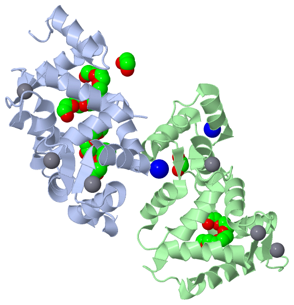
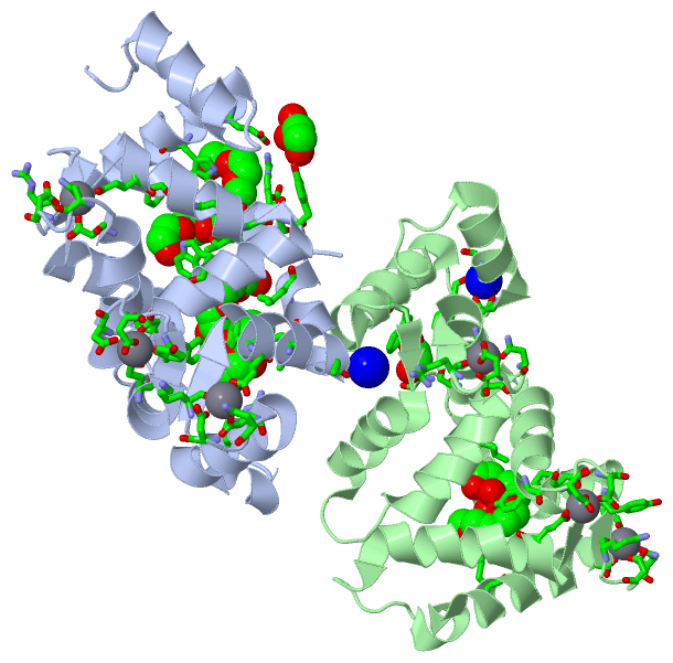
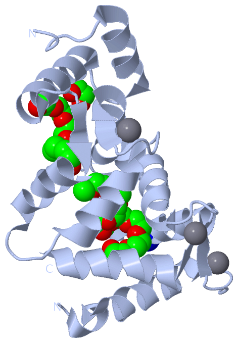
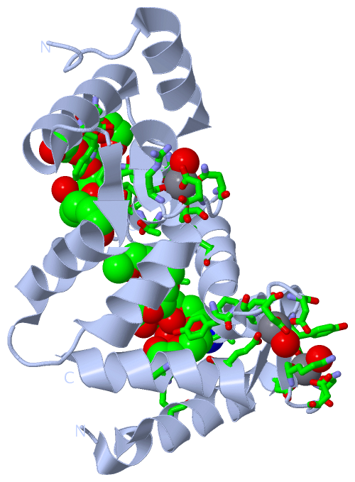
 Description