|
|
|
|
 Description
|
|
Compounds
|
||||||||||||||||||||||||||||||||||||||||||||
Chains, Units
Summary Information (see also Sequences/Alignments below) |
Ligands, Modified Residues, Ions (0, 0)| (no "Ligand,Modified Residues,Ions" information available for 1FC9) |
Sites (0, 0)| (no "Site" information available for 1FC9) |
SS Bonds (1, 1)
Asymmetric/Biological Unit
|
||||||||
Cis Peptide Bonds (0, 0)| (no "Cis Peptide Bond" information available for 1FC9) |
SAPs(SNPs)/Variants (0, 0)| (no "SAP(SNP)/Variant" information available for 1FC9) |
PROSITE Motifs (1, 1)
Asymmetric/Biological Unit (1, 1)
|
||||||||||||||||||||||||
Exons (0, 0)| (no "Exon" information available for 1FC9) |
Sequences/Alignments
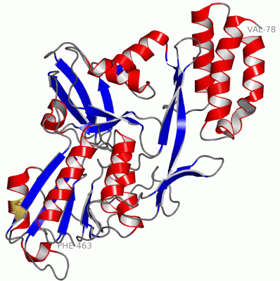
Asymmetric/Biological UnitChain A from PDB Type:PROTEIN Length:386 aligned with CTPA_TETOB | O04073 from UniProtKB/Swiss-Prot Length:464 Alignment length:386 87 97 107 117 127 137 147 157 167 177 187 197 207 217 227 237 247 257 267 277 287 297 307 317 327 337 347 357 367 377 387 397 407 417 427 437 447 457 CTPA_TETOB 78 VTSEQLLFLEAWRAVDRAYVDKSFNGQSWFKLRETYLKKEPMDRRAQTYDAIRKLLAVLDDPFTRFLEPSRLAALRRGTAGSVTGVGLEITYDGGSGKDVVVLTPAPGGPAEKAGARAGDVIVTVDGTAVKGLSLYDVSDLLQGEADSQVEVVLHAPGAPSNTRTLQLTRQKVTINPVTFTTCSNVAAAALPPGAAKQQLGYVRLATFNSNTTAAAQQAFTELSKQGVAGLVLDIRNNGGGLFPAGVNVARMLVDRGDLVLIADSQGIRDIYSADGNSIDSATPLVVLVNRGTASASEVLAGALKDSKRGLIAGERTFGKGLIQTVVDLSDGSGVAVTVARYQTPAGVDINKIGVSPDVQLDPEVLPTDLEGVCRVLGSDAAPRLF 463 SCOP domains d1fc9a4 A:78-156,A:249-463 Photosystem II D1 C-terminal processing protease d1fc9a3 A:157-248 Photosystem II D1 C-terminal processing protease d1fc9a4 A:78-156,A:249-463 Photosystem II D1 C-terminal processing protease SCOP domains CATH domains 1fc9A01 A:78-160,A:400-414 [code=3.30.750.34, no name defined] 1fc9A02 A:161-252 [code=2.30.42.10, no name defined] 1fc9A03 A:253-399,A:415-463 2-enoyl-CoA Hydratase; Chain A, domain 1 1fc9A01 1fc9A03 A:253-399,A:415-463 CATH domains Pfam domains -------------------------------------------------------------------------------------------------------------------------------------------------------------------------------------------------------------------------------------------------------------------------------------------------------------------------------------------------------------------------------------------------- Pfam domains SAPs(SNPs) -------------------------------------------------------------------------------------------------------------------------------------------------------------------------------------------------------------------------------------------------------------------------------------------------------------------------------------------------------------------------------------------------- SAPs(SNPs) PROSITE -----------------------------------------------------------------------PDZ PDB: A:149-220 UniProt: 149-220 --------------------------------------------------------------------------------------------------------------------------------------------------------------------------------------------------------------------------------------------------- PROSITE Transcript -------------------------------------------------------------------------------------------------------------------------------------------------------------------------------------------------------------------------------------------------------------------------------------------------------------------------------------------------------------------------------------------------- Transcript 1fc9 A 78 VTSEQLLFLEAWRAVDRAYVDKSFNGQSWFKLRETYLKKEPMDRRAQTYDAIRKLLAVLDDPFTRFLEPSRLAALRRGTAGSVTGVGLEITYDGGSGKDVVVLTPAPGGPAEKAGARAGDVIVTVDGTAVKGLSLYDVSDLLQGEADSQVEVVLHAPGAPSNTRTLQLTRQKVTINPVTFTTCSNVAAAALPPGAAKQQLGYVRLATFNSNTTAAAQQAFTELSKQGVAGLVLDIRNNGGGLFPAGVNVARMLVDRGDLVLIADSQGIRDIYSADGNSIDSATPLVVLVNRGTASASEVLAGALKDSKRGLIAGERTFGKGLIQTVVDLSDGSGVAVTVARYQTPAGVDINKIGVSPDVQLDPEVLPTDLEGVCRVLGSDAAPRLF 463 87 97 107 117 127 137 147 157 167 177 187 197 207 217 227 237 247 257 267 277 287 297 307 317 327 337 347 357 367 377 387 397 407 417 427 437 447 457
|
||||||||||||||||||||
SCOP Domains (2, 2)
Asymmetric/Biological Unit
 |
CATH Domains (3, 3)
Asymmetric/Biological Unit
|
Pfam Domains (0, 0)| (no "Pfam Domain" information available for 1FC9) |
Gene Ontology (8, 8)|
Asymmetric/Biological Unit(hide GO term definitions) Chain A (CTPA_TETOB | O04073)
|
||||||||||||||||||||||||||||||||||||||||||||||||||||||||||||||||||
Interactive Views
|
||||||||||||||||||||||||||||||||||||||||||||||||||||||||||||||||||||||||||||||||||||||||||||||||||||||||||||||||||||
Still Images
|
||||||||||||||||
Databases
|
||||||||||||||||||||||||||||||||||||||||||||||||||||||||||||||||||||||||||||||||||||||||||||||||||||||||||||||||||||||||||||||||||||||||||||||||||||||||||||||||
Analysis Tools
|
|||||||||||||||||||||||||||||||||||||||||||||||||||||||||||||
Entries Sharing at Least One Protein Chain (UniProt ID)
Related Entries Specified in the PDB File
|
|