|
|
|
|
 Description
|
|
Compounds
|
||||||||||||||||||||||||||||
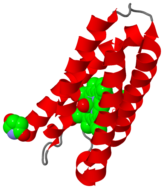
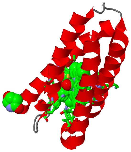
Chains, Units
Summary Information (see also Sequences/Alignments below) |
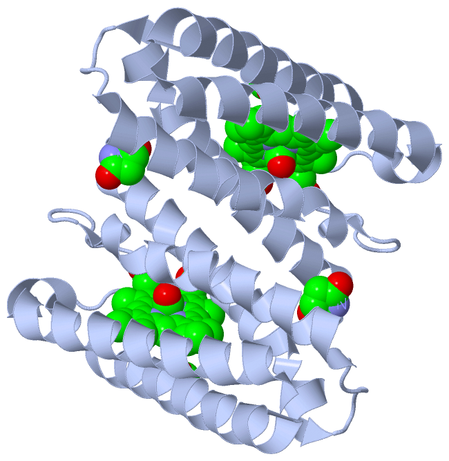
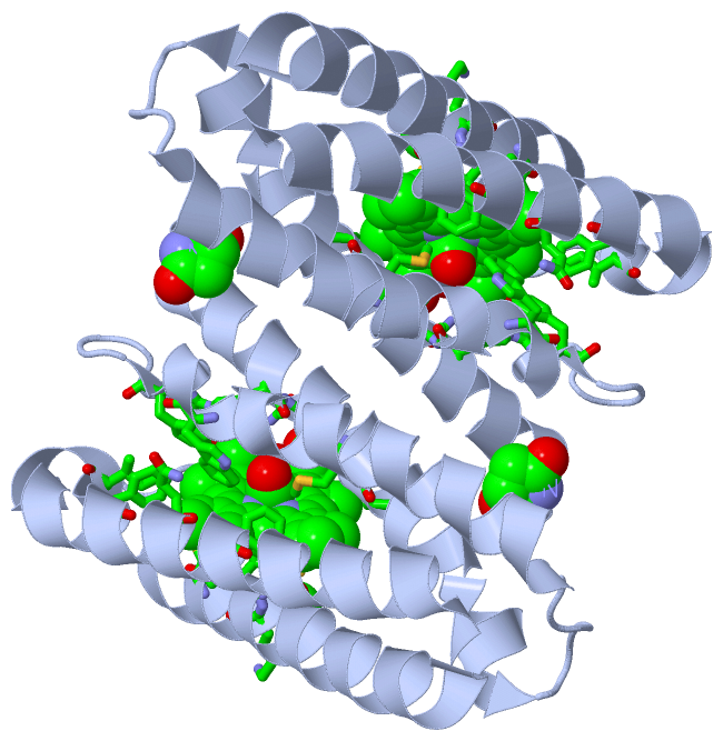
Ligands, Modified Residues, Ions (3, 4)| Asymmetric Unit (3, 4) Biological Unit 1 (3, 8) |
Sites (3, 3)
Asymmetric Unit (3, 3)
|
SS Bonds (0, 0)| (no "SS Bond" information available for 1E86) |
Cis Peptide Bonds (0, 0)| (no "Cis Peptide Bond" information available for 1E86) |
SAPs(SNPs)/Variants (0, 0)| (no "SAP(SNP)/Variant" information available for 1E86) |
PROSITE Motifs (1, 1)
Asymmetric Unit (1, 1)
|
||||||||||||||||||||||||||||||||||||||||||||||||
Exons (0, 0)| (no "Exon" information available for 1E86) |
Sequences/Alignments
Asymmetric UnitChain A from PDB Type:PROTEIN Length:126 aligned with CYCP_ALCXX | P00138 from UniProtKB/Swiss-Prot Length:127 Alignment length:126 10 20 30 40 50 60 70 80 90 100 110 120 CYCP_ALCXX 1 QFAKPEDAVKYRQSALTLMASHFGRMTPVVKGQAPYDAAQIKANVEVLKTLSALPWAAFGPGTEGGDARPEIWSDAASFKQKQQAFQDNIVKLSAAADAGDLDKLRAAFGDVGASCKACHDAYRKK 126 SCOP domains d1e86a_ A: Cytochrome c' SCOP domains CATH domains -1e86A00 A:2-126 [code=1.20.120.10, no name defined] CATH domains Pfam domains ------------------------------------------------------------------------------------------------------------------------------ Pfam domains SAPs(SNPs) ------------------------------------------------------------------------------------------------------------------------------ SAPs(SNPs) PROSITE ------CYTCII PDB: A:7-125 UniProt: 7-125 - PROSITE Transcript ------------------------------------------------------------------------------------------------------------------------------ Transcript 1e86 A 1 xFAKPEDAVKYRQSALTLMASHFGRMTPVVKGQAPYDAAQIKANVEVLKTLSALPWAAFGPGTEGGDARPEIWSDAASFKQKQQAFQDNIVKLSAAADAGDLDKLRAAFGDVGASCKACHDAYRKK 126 | 10 20 30 40 50 60 70 80 90 100 110 120 | 1-PCA
|
||||||||||||||||||||
SCOP Domains (1, 1)
Asymmetric Unit

|
CATH Domains (1, 1)
Asymmetric Unit
|
Pfam Domains (0, 0)| (no "Pfam Domain" information available for 1E86) |
Gene Ontology (7, 7)|
Asymmetric Unit(hide GO term definitions) Chain A (CYCP_ALCXX | P00138)
|
||||||||||||||||||||||||||||||||||||||||||||||||||||||||||||
Interactive Views
|
||||||||||||||||||||||||||||||||||||||||||||||||||||||||||||||||||||||||||||||||||||||||||||||||||||||||||||||||||||||||||||||||||||||||||||||||||||||||||||||||||||
Still Images
|
||||||||||||||||
Databases
|
||||||||||||||||||||||||||||||||||||||||||||||||||||||||||||||||||||||||||||||||||||||||||||||||||||||||||||||||||||||||||||||||||||||||||||||||||||||||||||||||
Analysis Tools
|
|||||||||||||||||||||||||||||||||||||||||||||||||||||||||||||
Entries Sharing at Least One Protein Chain (UniProt ID)
Related Entries Specified in the PDB File
|
|