Show PDB file:   
         Plain Text   HTML   (compressed file size)
QuickSearch:   
by PDB,NDB,UniProt,PROSITE Code or Search Term(s)  
(-)NMR Structure - manually
(-)NMR Structure - model 1
(-)NMR Structure - model 1, sites
(-)NMR Structure - all models
collapse expand < >
Image NMR Structure - manually
NMR Structure - manually  (Jmol Viewer)
Image NMR Structure - model 1
NMR Structure - model 1  (Jmol Viewer)
Image NMR Structure - model 1, sites
NMR Structure - model 1, sites  (Jmol Viewer)
Image NMR Structure - all models
NMR Structure - all models  (Jmol Viewer)

(-) Description

Title :  THREE-DIMENSIONAL STRUCTURE OF THE APO FORM OF THE N-TERMINAL EGF-LIKE MODULE OF BLOOD COAGULATION FACTOR X AS DETERMINED BY NMR SPECTROSCOPY AND SIMULATED FOLDING
 
Authors :  M. Ullner, M. Selander, E. Persson, J. Stenflo, T. Drakenberg, O. Teleman
Date :  21 Apr 92  (Deposition) - 31 Jan 94  (Release) - 24 Feb 09  (Revision)
Method :  SOLUTION NMR
Resolution :  NOT APPLICABLE
Chains :  NMR Structure  :  A  (13x)
Keywords :  Coagulation Factor (Keyword Search: [Gene Ontology, PubMed, Web (Google))
 
Reference :  M. Ullner, M. Selander, E. Persson, J. Stenflo, T. Drakenberg, O. Teleman
Three-Dimensional Structure Of The Apo Form Of The N-Terminal Egf-Like Module Of Blood Coagulation Factor X As Determined By Nmr Spectroscopy And Simulated Folding.
Biochemistry V. 31 5974 1992
PubMed-ID: 1627540  |  Reference-DOI: 10.1021/BI00141A004
(for further references see the PDB file header)

(-) Compounds

Molecule 1 - EGF-LIKE MODULE OF BLOOD COAGULATION FACTOR X
    ChainsA
    EngineeredYES
    Organism CommonCATTLE
    Organism ScientificBOS TAURUS
    Organism Taxid9913

 Structural Features

(-) Chains, Units

  
NMR Structure (13x)

Summary Information (see also Sequences/Alignments below)

(-) Ligands, Modified Residues, Ions  (1, 1)

NMR Structure (1, 1)
No.NameCountTypeFull Name
1OH1Ligand/IonHYDROXIDE ION

(-) Sites  (1, 1)

NMR Structure (1, 1)
No.NameEvidenceResiduesDescription
1AC1SOFTWAREASP A:63 , GLY A:64BINDING SITE FOR RESIDUE OH A 87

(-) SS Bonds  (3, 3)

NMR Structure
No.Residues
1A:50 -A:61
2A:55 -A:70
3A:72 -A:81

(-) Cis Peptide Bonds  (0, 0)

(no "Cis Peptide Bond" information available for 1APO)

 Sequence-Structure Mapping

(-) SAPs(SNPs)/Variants  (0, 0)

(no "SAP(SNP)/Variant" information available for 1APO)

(-) PROSITE Motifs  (5, 4)

NMR Structure (5, 4)
 PROSITEUniProtKBPDB
No.IDACDescriptionIDLocationCountLocation
1EGF_3PS50026 EGF-like domain profile.FA10_BOVIN86-122  1A:46-82
2EGF_CAPS01187 Calcium-binding EGF-like domain signature.FA10_BOVIN86-110  1A:46-70
3ASX_HYDROXYLPS00010 Aspartic acid and asparagine hydroxylation site.FA10_BOVIN101-112  1A:61-72
4EGF_1PS00022 EGF-like domain signature 1.FA10_BOVIN110-121  1A:70-81
5EGF_2PS01186 EGF-like domain signature 2.FA10_BOVIN110-121
149-164
  1A:70-81
-

(-) Exons   (3, 3)

NMR Structure (3, 3)
 ENSEMBLUniProtKBPDB
No.Transcript IDExonExon IDGenome LocationLengthIDLocationLengthCountLocationLength
1.1ENSBTAT000000217891ENSBTAE00000377900chr12:84664833-8466490270FA10_BOVIN1-24240--
1.2ENSBTAT000000217892ENSBTAE00000278163chr12:84666990-84667150161FA10_BOVIN24-77540--
1.3ENSBTAT000000217893ENSBTAE00000177940chr12:84671119-8467114325FA10_BOVIN78-8691A:45-462
1.4ENSBTAT000000217894ENSBTAE00000177941chr12:84672093-84672206114FA10_BOVIN86-124391A:46-8439
1.5ENSBTAT000000217895ENSBTAE00000425962chr12:84673472-84673603132FA10_BOVIN124-168451A:84-863
1.6ENSBTAT000000217896ENSBTAE00000408790chr12:84675374-84675615242FA10_BOVIN168-248810--
1.7ENSBTAT000000217897ENSBTAE00000412504chr12:84676748-84676865118FA10_BOVIN249-288400--
1.8ENSBTAT000000217898ENSBTAE00000414972chr12:84677559-84678217659FA10_BOVIN288-4922050--

(-) Sequences/Alignments

NMR Structure
   Reformat: Number of residues per line =  ('0' or empty: single-line sequence representation)
  Number of residues per labelling interval =   
  UniProt sequence: complete  aligned part    
   Show mapping: SCOP domains CATH domains Pfam domains Secondary structure (by author)
SAPs(SNPs) PROSITE motifs Exons
(details for a mapped element are shown in a popup box when the mouse pointer rests over it)
Chain A from PDB  Type:PROTEIN  Length:42
 aligned with FA10_BOVIN | P00743 from UniProtKB/Swiss-Prot  Length:492

    Alignment length:42
                                    94       104       114       124  
           FA10_BOVIN    85 KDGDQCEGHPCLNQGHCKDGIGDYTCTCAEGFEGKNCEFSTR 126
               SCOP domains d1apoa_ A: Factor X, N-terminal module     SCOP domains
               CATH domains 1apoA00 A:45-86 Laminin                    CATH domains
               Pfam domains ------------------------------------------ Pfam domains
         Sec.struct. author ..............eeeee....eeeee...ee.....ee.. Sec.struct. author
                 SAPs(SNPs) ------------------------------------------ SAPs(SNPs)
                PROSITE (1) -EGF_CA  PDB: A:46-70     ---------------- PROSITE (1)
                PROSITE (2) -EGF_3  PDB: A:46-82 UniProt: 86-122  ---- PROSITE (2)
                PROSITE (3) ----------------ASX_HYDROXYL-------------- PROSITE (3)
                PROSITE (4) -------------------------EGF_2       ----- PROSITE (4)
                PROSITE (5) -------------------------EGF_1       ----- PROSITE (5)
           Transcript 1 (1) 1.-------------------------------------1.5 Transcript 1 (1)
           Transcript 1 (2) -Exon 1.4  PDB: A:46-84 UniProt: 86-124 -- Transcript 1 (2)
                 1apo A  45 KDGDQCEGHPCLNQGHCKDGIGDYTCTCAEGFEGKNCEFSTR  86
                                    54        64        74        84  

   Legend:   → Mismatch (orange background)
  - → Gap (green background, '-', border residues have a numbering label)
    → Modified Residue (blue background, lower-case, 'x' indicates undefined single-letter code, labelled with number + name)
  x → Chemical Group (purple background, 'x', labelled with number + name, e.g. ACE or NH2)
  extra numbering lines below/above indicate numbering irregularities and modified residue names etc., number ends below/above '|'

 Classification and Annotation

(-) SCOP Domains  (1, 1)

NMR Structure

(-) CATH Domains  (1, 1)

NMR Structure
(-)
Class: Mainly Beta (13760)

(-) Pfam Domains  (0, 0)

(no "Pfam Domain" information available for 1APO)

(-) Gene Ontology  (11, 11)

NMR Structure(hide GO term definitions)
Chain A   (FA10_BOVIN | P00743)
molecular function
    GO:0005509    calcium ion binding    Interacting selectively and non-covalently with calcium ions (Ca2+).
    GO:0016787    hydrolase activity    Catalysis of the hydrolysis of various bonds, e.g. C-O, C-N, C-C, phosphoric anhydride bonds, etc. Hydrolase is the systematic name for any enzyme of EC class 3.
    GO:0008233    peptidase activity    Catalysis of the hydrolysis of a peptide bond. A peptide bond is a covalent bond formed when the carbon atom from the carboxyl group of one amino acid shares electrons with the nitrogen atom from the amino group of a second amino acid.
    GO:0005543    phospholipid binding    Interacting selectively and non-covalently with phospholipids, a class of lipids containing phosphoric acid as a mono- or diester.
    GO:0004252    serine-type endopeptidase activity    Catalysis of the hydrolysis of internal, alpha-peptide bonds in a polypeptide chain by a catalytic mechanism that involves a catalytic triad consisting of a serine nucleophile that is activated by a proton relay involving an acidic residue (e.g. aspartate or glutamate) and a basic residue (usually histidine).
    GO:0008236    serine-type peptidase activity    Catalysis of the hydrolysis of peptide bonds in a polypeptide chain by a catalytic mechanism that involves a catalytic triad consisting of a serine nucleophile that is activated by a proton relay involving an acidic residue (e.g. aspartate or glutamate) and a basic residue (usually histidine).
biological process
    GO:0007596    blood coagulation    The sequential process in which the multiple coagulation factors of the blood interact, ultimately resulting in the formation of an insoluble fibrin clot; it may be divided into three stages: stage 1, the formation of intrinsic and extrinsic prothrombin converting principle; stage 2, the formation of thrombin; stage 3, the formation of stable fibrin polymers.
    GO:0007599    hemostasis    The stopping of bleeding (loss of body fluid) or the arrest of the circulation to an organ or part.
    GO:0051897    positive regulation of protein kinase B signaling    Any process that activates or increases the frequency, rate or extent of protein kinase B signaling, a series of reactions mediated by the intracellular serine/threonine kinase protein kinase B.
    GO:0006508    proteolysis    The hydrolysis of proteins into smaller polypeptides and/or amino acids by cleavage of their peptide bonds.
cellular component
    GO:0005576    extracellular region    The space external to the outermost structure of a cell. For cells without external protective or external encapsulating structures this refers to space outside of the plasma membrane. This term covers the host cell environment outside an intracellular parasite.
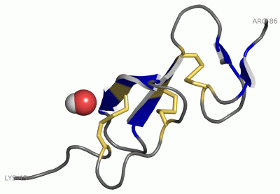
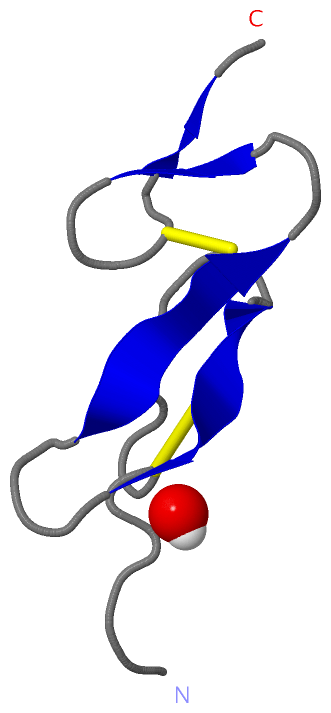
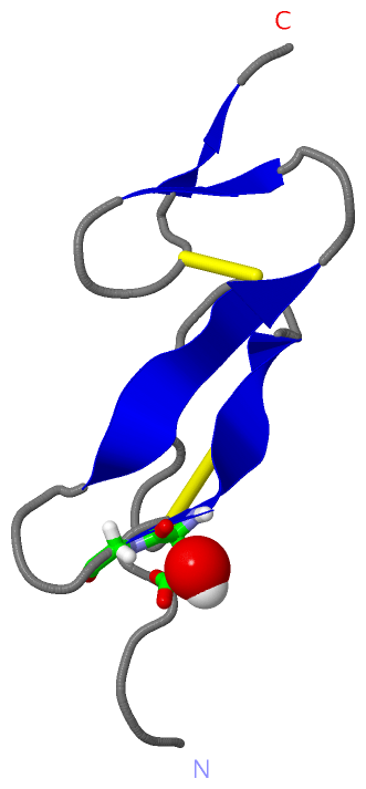
 Visualization

(-) Interactive Views

NMR Structure
  Complete Structure
    Jena3D(integrated viewing of ligand, site, SAP, PROSITE, SCOP information)
    WebMol | AstexViewer[tm]@PDBe
(Java Applets, require no local installation except for Java; loading may be slow)
    STRAP
(Java WebStart application, automatic local installation, requires Java; full application with system access!)
    RasMol
(require local installation)
    Molscript (VRML)
(requires installation of a VRML viewer; select preferred view via VRML and generate a mono or stereo PDF format file)
 
  Ligands, Modified Residues, Ions
    OH  [ RasMol | Jena3D ]  +environment [ RasMol | Jena3D ]
 
  Sites
    AC1  [ RasMol ]  +environment [ RasMol ]
 
  Cis Peptide Bonds
(no "Cis Peptide Bonds" information available for 1apo)
 

(-) Still Images

Jmol
  protein: cartoon or spacefill or dots and stick; nucleic acid: cartoon and stick; ligands: spacefill; active site: stick
Molscript
  protein, nucleic acid: cartoon; ligands: spacefill; active site: ball and stick

 Databases and Analysis Tools

(-) Databases

Access by PDB/NDB ID
  1apo
    Family and Domain InformationProDom | SYSTERS
    General Structural InformationGlycoscienceDB | MMDB | NDB | OCA | PDB | PDBe | PDBj | PDBsum | PDBWiki | PQS | PROTEOPEDIA
    Orientation in MembranesOPM
    Protein SurfaceSURFACE
    Secondary StructureDSSP (structure derived) | HSSP (homology derived)
    Structural GenomicsGeneCensus
    Structural NeighboursCE | VAST
    Structure ClassificationCATH | Dali | SCOP
    Validation and Original DataBMRB Data View | BMRB Restraints Grid | EDS | PROCHECK | RECOORD | WHAT_CHECK
 
Access by UniProt ID/Accession number
  FA10_BOVIN | P00743
    Comparative Protein Structure ModelsModBase
    Genomic InformationEnsembl
    Protein-protein InteractionDIP
    Sequence, Family and Domain InformationInterPro | Pfam | SMART | UniProtKB/SwissProt
 
Access by Enzyme Classificator   (EC Number)
  (no 'Enzyme Classificator' available)
    General Enzyme InformationBRENDA | EC-PDB | Enzyme | IntEnz
    PathwayKEGG | MetaCyc
 
Access by Disease Identifier   (MIM ID)
  (no 'MIM ID' available)
    Disease InformationOMIM
 
Access by GenAge ID
  (no 'GenAge ID' available)
    Age Related InformationGenAge

(-) Analysis Tools

Access by PDB/NDB ID
    Domain InformationXDom
    Interatomic Contacts of Structural UnitsCSU
    Ligand-protein ContactsLPC
    Protein CavitiescastP
    Sequence and Secondary StructurePDBCartoon
    Structure AlignmentSTRAP(Java WebStart application, automatic local installation, requires Java; full application with system access!)
    Structure and Sequence BrowserSTING
 
Access by UniProt ID/Accession number
  FA10_BOVIN | P00743
    Protein Disorder PredictionDisEMBL | FoldIndex | GLOBPLOT (for more information see DisProt)

 Related Entries

(-) Entries Sharing at Least One Protein Chain (UniProt ID)

UniProtKB/Swiss-Prot
        FA10_BOVIN | P007431ccf 1iod 1kig 1whe 1whf

(-) Related Entries Specified in the PDB File

(no "Related Entries Specified in the PDB File" available for 1APO)