|
|
 Atom Coloring Scheme
| Atom | Color | RGB Code |
| Oxygen | Red | 255, 0, 0 |
| Nitrogen | Blue | 0, 0, 255 |
| Carbon | Green | 0, 255, 0 |
| Phosphorus | Magenta | 255, 0, 0 |
| Sulfur | Yellow | 255, 255, 0 |
This scheme is adopted for almost all images generated with RasMol, InsightII, WebLab and MolScript. There are, however, a few MolMol, Setor and Sybyl images, where carbon is colored gray and phosphorus is not colored magenta .
ATP-gammaS spacefill representation with MolMol.
Residue Coloring SchemeResidues are color-coded according to the scheme originally used by the program SETOR written by Stephen Evans.
Residue coloring scheme.
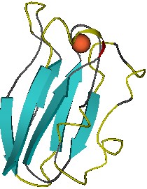
Secondary Structure Elements Coloring and DrawingAlpha-helices are either drawn as cylinders or as ribbons. The preferred color used in most cases is red (RasMol, InsightII, WebLab). MolScript and MolMol draw helices as ribbons with a different outside (red) and inside (yellow) coloring. Beta-strands are colored blue (RasMol, InsightII, WebLab) or cyan (MolScript, MolMol). A few graphics programs color turns in yellow (MolScript, RasMol, InsightII).
The MHC class I binding groove, built by alpha-helices and a beta-sheet on the bottom, is shown complexed with a stretched peptide (MolMol image from part of the MHC structure, PDB code: 1A1N).
Secondary Structure DeterminationSecondary structure information is often but not always included with the coordinate files from the NDB and PDB. RasMol, MolMol, InsightII, Midas and Setor use the PDB secondary structure information if there is any. If there are no secondary structure information records provided with the coordinate file, then the secondary structure assignment is calculated according to the Kabsch and Sander DSSP algorithm. MolScript and WebLab do not use the PDB secondary structure information at all, but calculate this information in all cases. Moreover, some programs cannot display very short helices (3 amino acids).
Warning: This may lead to slightly different secondary structure representations for one and the same structure .
|
 |
Different secondary structure determination (MolMol and MolScript images of plastocyanin, PDB code: 1PLC)
Active Sites and Disulfide BondsSites as defined in the coordinate file are displayed as ball and sticks by MolScript or sticks only by RasMol and MolMol.
Disulfide bonds are displayed if there are SS bond records in the coordinate file. They are shown as lines by MolScript, RasMol or MolMol. However, older MolMol representations do not display SS bonds. SS bonds are often not shown in InsightII, Midas and WebLab images. Setor and Prepi adopt a cartoon-like representation of SS bonds.
The trypsin catalytic site amino acids His57 , Asp102 and Ser195 are shown as thick sticks, the thin lines are disulfide bonds (image generated with MolMol, PDB code: 1SGT).
Ligands, Modified Residues, IonsThe heterogen section of the PDB file header defines 'non-standard' residues, such as modified amino acids or nucleotides, prosthetic groups, inhibitors, metal ions and solvent components. Non-standard residues are shown as spacefill representation in RasMol, MolScript and MolMol images . For more detailed information see the PDB Contents Guide (PDF format). The nucleotide I (inosine) and the modified nucleotides +C, +G, +A, +T, +U, +I are considered as standard residues according to the PDB definition. They are, however, treated in a different way by RasMol and MolScript. RasMol displays these nucleotides as sticks and in MolScript images the base plates are drawn in the same color as the "mother" nucleotide but contrary to C, G, A, T, U, I the lines are colored in an atom-specific manner.
|
|
|
|
Beutenbergstraße 11 D-07745 Jena • Germany |
Phone: +49 3641 65-6000 Fax: +49 3641 65-6351 |
E-mail: info@leibniz-fli.de www.leibniz-fli.de |
Data Privacy Imprint |
 |
