Asymmetric Unit (10, 10)
| No. | Name | Evidence | Residues | Description |
|---|
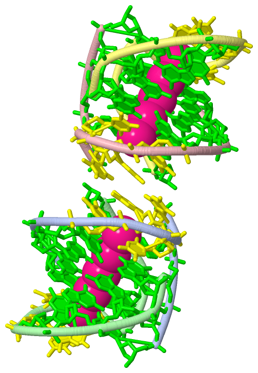
| 01 | AC1 | SOFTWARE | DG A:1001 , DG A:1012 , K A:5016 , DG B:2004 , DT B:2005 , DT B:2007 , DG B:2009 , HOH B:6004 | BINDING SITE FOR RESIDUE K B 5013 |
| 02 | AC2 | SOFTWARE | DG A:1002 , DG A:1003 , DG A:1010 , DG A:1011 , K A:5016 , DG B:2002 , DG B:2003 , DG B:2010 , DG B:2011 , K B:5015 | BINDING SITE FOR RESIDUE K B 5014 |
| 03 | AC3 | SOFTWARE | DG A:1003 , DG A:1004 , DG A:1009 , DG A:1010 , K A:5017 , DG B:2001 , DG B:2002 , DG B:2011 , DG B:2012 , K B:5014 | BINDING SITE FOR RESIDUE K B 5015 |
| 04 | AC4 | SOFTWARE | DG A:1001 , DG A:1002 , DG A:1011 , DG A:1012 , DG B:2003 , DG B:2004 , DG B:2009 , DG B:2010 , K B:5013 , K B:5014 | BINDING SITE FOR RESIDUE K A 5016 |
| 05 | AC5 | SOFTWARE | DG A:1004 , DT A:1005 , DT A:1007 , DG A:1009 , DG B:2001 , DG B:2012 , K B:5015 | BINDING SITE FOR RESIDUE K A 5017 |
| 06 | AC6 | SOFTWARE | DG C:3002 , DG C:3003 , DG C:3010 , DG C:3011 , K C:5019 , K C:5020 , DG D:4002 , DG D:4003 , DG D:4010 , DG D:4011 | BINDING SITE FOR RESIDUE K C 5018 |
| 07 | AC7 | SOFTWARE | DG C:3001 , DG C:3002 , DG C:3011 , DG C:3012 , K C:5018 , DG D:4003 , DG D:4004 , DG D:4009 , DG D:4010 , K D:5021 | BINDING SITE FOR RESIDUE K C 5019 |
| 08 | AC8 | SOFTWARE | DG C:3003 , DG C:3004 , DG C:3009 , DG C:3010 , K C:5018 , K C:5022 , DG D:4001 , DG D:4002 , DG D:4011 , DG D:4012 | BINDING SITE FOR RESIDUE K C 5020 |
| 09 | AC9 | SOFTWARE | DG C:3001 , DG C:3012 , K C:5019 , DG D:4004 , DT D:4005 , DT D:4007 , DG D:4009 | BINDING SITE FOR RESIDUE K D 5021 |
| 10 | BC1 | SOFTWARE | DG C:3004 , DT C:3005 , DT C:3007 , DG C:3009 , K C:5020 , DG D:4001 , DG D:4012 | BINDING SITE FOR RESIDUE K C 5022 |
|
 Description