Show PDB file:   
         Plain Text   HTML   (compressed file size)
QuickSearch:   
by PDB,NDB,UniProt,PROSITE Code or Search Term(s)  
(-)NMR Structure - manually
(-)NMR Structure - model 1
(-)NMR Structure - model 1, sites
(-)NMR Structure - all models
collapse expand < >
Image NMR Structure - manually
NMR Structure - manually  (Jmol Viewer)
Image NMR Structure - model 1
NMR Structure - model 1  (Jmol Viewer)
Image NMR Structure - model 1, sites
NMR Structure - model 1, sites  (Jmol Viewer)
Image NMR Structure - all models
NMR Structure - all models  (Jmol Viewer)

(-) Description

Title :  SOLUTION NMR STRUCTURE OF THE L22V MUTANT DNA BINDING DOMAIN OF AREA COMPLEXED TO A 13 BP DNA CONTAINING A TGATA SITE, 34 STRUCTURES
 
Authors :  G. M. Clore, M. Starich, M. Wikstrom, A. M. Gronenborn
Date :  07 Nov 97  (Deposition) - 28 Jan 98  (Release) - 24 Feb 09  (Revision)
Method :  SOLUTION NMR
Resolution :  NOT APPLICABLE
Chains :  NMR Structure  :  A,B,C  (34x)
Keywords :  Dna Binding Protein, Transcription Factor, Zinc Binding Domain, Complex (Transcription Regulation/Dna), Transcription/Dna Complex (Keyword Search: [Gene Ontology, PubMed, Web (Google))
 
Reference :  M. R. Starich, M. Wikstrom, S. Schumacher, H. N. Arst Jr. , A. M. Gronenborn, G. M. Clore
The Solution Structure Of The Leu22-->Val Mutant Area Dna Binding Domain Complexed With A Tgatag Core Element Defines A Role For Hydrophobic Packing In The Determination Of Specificity.
J. Mol. Biol. V. 277 621 1998
PubMed-ID: 9533884  |  Reference-DOI: 10.1006/JMBI.1997.1626
(for further references see the PDB file header)

(-) Compounds

Molecule 1 - DNA (5'- D(*CP*AP*GP*TP*GP*AP*TP*AP*GP*AP*GP*AP*C)-3')
    ChainsB
    EngineeredYES
    SyntheticYES
 
Molecule 2 - DNA (5'- D(*GP*TP*CP*TP*CP*TP*AP*TP*CP*AP*CP*TP*G)-3')
    ChainsC
    EngineeredYES
    SyntheticYES
 
Molecule 3 - NITROGEN REGULATORY PROTEIN AREA
    ChainsA
    EngineeredYES
    Expression SystemESCHERICHIA COLI
    Expression System Taxid562
    FragmentDNA BINDING DOMAIN
    GenePOTENTIAL
    MutationYES
    Organism ScientificEMERICELLA NIDULANS
    Organism Taxid162425

 Structural Features

(-) Chains, Units

  
NMR Structure (34x)

Summary Information (see also Sequences/Alignments below)

(-) Ligands, Modified Residues, Ions  (1, 1)

NMR Structure (1, 1)
No.NameCountTypeFull Name
1ZN1Ligand/IonZINC ION

(-) Sites  (1, 1)

NMR Structure (1, 1)
No.NameEvidenceResiduesDescription
1AC1SOFTWARECYS A:12 , CYS A:15 , CYS A:33 , CYS A:36BINDING SITE FOR RESIDUE ZN A 67

(-) SS Bonds  (0, 0)

(no "SS Bond" information available for 7GAT)

(-) Cis Peptide Bonds  (0, 0)

(no "Cis Peptide Bond" information available for 7GAT)

 Sequence-Structure Mapping

(-) SAPs(SNPs)/Variants  (0, 0)

(no "SAP(SNP)/Variant" information available for 7GAT)

(-) PROSITE Motifs  (0, 0)

(no "PROSITE Motif" information available for 7GAT)

(-) Exons   (0, 0)

(no "Exon" information available for 7GAT)

(-) Sequences/Alignments

NMR Structure
   Reformat: Number of residues per line =  ('0' or empty: single-line sequence representation)
  Number of residues per labelling interval =   
  UniProt sequence: complete  aligned part    
   Show mapping: SCOP domains CATH domains Pfam domains Secondary structure (by author)
SAPs(SNPs) PROSITE motifs Exons
(details for a mapped element are shown in a popup box when the mouse pointer rests over it)
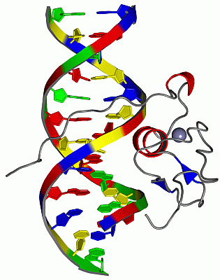
Chain A from PDB  Type:PROTEIN  Length:66
                                                                                                  
               SCOP domains d7gata_ A: Erythroid transcription factor GATA-1                   SCOP domains
               CATH domains 7gatA00 A:1-66 Erythroid Transcription Factor GATA-1, subunit A    CATH domains
               Pfam domains ------------------------------------------------------------------ Pfam domains
         Sec.struct. author .......................ee.....ee.hhhhhhhhhh.....hhh............... Sec.struct. author
                 SAPs(SNPs) ------------------------------------------------------------------ SAPs(SNPs)
                    PROSITE ------------------------------------------------------------------ PROSITE
                 Transcript ------------------------------------------------------------------ Transcript
                 7gat A   1 MKNGEQNGPTTCTNCFTQTTPVWRRNPEGQPLCNACGLFLKLHGVVRPLSLKTDVIKKRNRNSANS  66
                                    10        20        30        40        50        60      

Chain B from PDB  Type:DNA  Length:13
                                             
                 7gat B 101 CAGTGATAGAGAC 113
                                   110   

Chain C from PDB  Type:DNA  Length:13
                                             
                 7gat C 114 GTCTCTATCACTG 126
                                   123   

   Legend:   → Mismatch (orange background)
  - → Gap (green background, '-', border residues have a numbering label)
    → Modified Residue (blue background, lower-case, 'x' indicates undefined single-letter code, labelled with number + name)
  x → Chemical Group (purple background, 'x', labelled with number + name, e.g. ACE or NH2)
  extra numbering lines below/above indicate numbering irregularities and modified residue names etc., number ends below/above '|'

 Classification and Annotation

(-) SCOP Domains  (1, 1)

NMR Structure

(-) CATH Domains  (1, 1)

NMR Structure
(-)
Class: Alpha Beta (26913)

(-) Pfam Domains  (0, 0)

(no "Pfam Domain" information available for 7GAT)

(-) Gene Ontology  (24, 24)

NMR Structure(hide GO term definitions)

 Visualization

(-) Interactive Views

NMR Structure
  Complete Structure
    Jena3D(integrated viewing of ligand, site, SAP, PROSITE, SCOP information)
    WebMol | AstexViewer[tm]@PDBe
(Java Applets, require no local installation except for Java; loading may be slow)
    STRAP
(Java WebStart application, automatic local installation, requires Java; full application with system access!)
    RasMol
(require local installation)
    Molscript (VRML)
(requires installation of a VRML viewer; select preferred view via VRML and generate a mono or stereo PDF format file)
 
  Ligands, Modified Residues, Ions
    ZN  [ RasMol | Jena3D ]  +environment [ RasMol | Jena3D ]
 
  Sites
    AC1  [ RasMol ]  +environment [ RasMol ]
 
  Cis Peptide Bonds
(no "Cis Peptide Bonds" information available for 7gat)
 

(-) Still Images

Jmol
  protein: cartoon or spacefill or dots and stick; nucleic acid: cartoon and stick; ligands: spacefill; active site: stick
Molscript
  protein, nucleic acid: cartoon; ligands: spacefill; active site: ball and stick

 Databases and Analysis Tools

(-) Databases

Access by PDB/NDB ID
  7gat
    Family and Domain InformationProDom | SYSTERS
    General Structural InformationGlycoscienceDB | MMDB | NDB | OCA | PDB | PDBe | PDBj | PDBsum | PDBWiki | PQS | PROTEOPEDIA
    Orientation in MembranesOPM
    Protein SurfaceSURFACE
    Secondary StructureDSSP (structure derived) | HSSP (homology derived)
    Structural GenomicsGeneCensus
    Structural NeighboursCE | VAST
    Structure ClassificationCATH | Dali | SCOP
    Validation and Original DataBMRB Data View | BMRB Restraints Grid | EDS | PROCHECK | RECOORD | WHAT_CHECK
 
Access by UniProt ID/Accession number
  AREA_EMENI | P17429
    Comparative Protein Structure ModelsModBase
    Genomic InformationEnsembl
    Protein-protein InteractionDIP
    Sequence, Family and Domain InformationInterPro | Pfam | SMART | UniProtKB/SwissProt
 
Access by Enzyme Classificator   (EC Number)
  (no 'Enzyme Classificator' available)
    General Enzyme InformationBRENDA | EC-PDB | Enzyme | IntEnz
    PathwayKEGG | MetaCyc
 
Access by Disease Identifier   (MIM ID)
  (no 'MIM ID' available)
    Disease InformationOMIM
 
Access by GenAge ID
  (no 'GenAge ID' available)
    Age Related InformationGenAge

(-) Analysis Tools

Access by PDB/NDB ID
    Domain InformationXDom
    Interatomic Contacts of Structural UnitsCSU
    Ligand-protein ContactsLPC
    Protein CavitiescastP
    Sequence and Secondary StructurePDBCartoon
    Structure AlignmentSTRAP(Java WebStart application, automatic local installation, requires Java; full application with system access!)
    Structure and Sequence BrowserSTING
 
Access by UniProt ID/Accession number
  AREA_EMENI | P17429
    Protein Disorder PredictionDisEMBL | FoldIndex | GLOBPLOT (for more information see DisProt)

 Related Entries

(-) Entries Sharing at Least One Protein Chain (UniProt ID)

UniProtKB/Swiss-Prot
        AREA_EMENI | P174292vus 2vut 2vuu 4gat 5gat 6gat

(-) Related Entries Specified in the PDB File

6gat REGULARIZED MEAN STRUCTURE