|
|
|
|
 Description
|
|
Compounds
|
||||||||||||||||||||||||||||||||||||||||||||
Chains, Units
Summary Information (see also Sequences/Alignments below) |
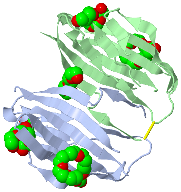
Ligands, Modified Residues, Ions (3, 7)| Asymmetric/Biological Unit (3, 7) |
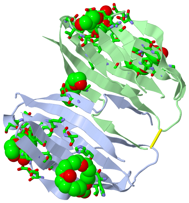
Sites (7, 7)
Asymmetric Unit (7, 7)
|
SS Bonds (1, 1)
Asymmetric/Biological Unit
|
||||||||
Cis Peptide Bonds (0, 0)| (no "Cis Peptide Bond" information available for 5XAW) |
SAPs(SNPs)/Variants (0, 0)| (no "SAP(SNP)/Variant" information available for 5XAW) |
PROSITE Motifs (0, 0)| (no "PROSITE Motif" information available for 5XAW) |
Exons (0, 0)| (no "Exon" information available for 5XAW) |
Sequences/Alignments
Asymmetric/Biological Unit
Chain A from PDB Type:PROTEIN Length:112
SCOP domains ---------------------------------------------------------------------------------------------------------------- SCOP domains
CATH domains ---------------------------------------------------------------------------------------------------------------- CATH domains
Pfam domains ---------------------------------------------------------------------------------------------------------------- Pfam domains
SAPs(SNPs) ---------------------------------------------------------------------------------------------------------------- SAPs(SNPs)
PROSITE ---------------------------------------------------------------------------------------------------------------- PROSITE
Transcript ---------------------------------------------------------------------------------------------------------------- Transcript
5xaw A 30 SLEVSVGKATDIYAVNGTEILLPCTFSSCFGFEDLHFRWTYNSSDAFKILIEGTVKNEKSDPKVTLKDDDRITLVNNISIVLRDLEFSDTGKYTCHVKNPLQHHATIFLQVV 152
39 49 59 69 79 89 99 || 116 126 136| 150
104| 136|
112 141
Chain B from PDB Type:PROTEIN Length:110
SCOP domains -------------------------------------------------------------------------------------------------------------- SCOP domains
CATH domains -------------------------------------------------------------------------------------------------------------- CATH domains
Pfam domains -------------------------------------------------------------------------------------------------------------- Pfam domains
SAPs(SNPs) -------------------------------------------------------------------------------------------------------------- SAPs(SNPs)
PROSITE -------------------------------------------------------------------------------------------------------------- PROSITE
Transcript -------------------------------------------------------------------------------------------------------------- Transcript
5xaw B 30 SLEVSVGKATDIYAVNGTEILLPCTFSSCFGFEDLHFRWTYNSSDAFKILIEGTVKNEKSDPKVTLKDDDRITLVNNISIVLRDLEFSDTGKYTCHVKNQHHATIFLQVV 152
39 49 59 69 79 89 99 || 116 126 142 152
104| 135|
112 142
|
||||||||||||||||||||
SCOP Domains (0, 0)| (no "SCOP Domain" information available for 5XAW) |
CATH Domains (0, 0)| (no "CATH Domain" information available for 5XAW) |
Pfam Domains (0, 0)| (no "Pfam Domain" information available for 5XAW) |
Gene Ontology (26, 26)|
Asymmetric/Biological Unit(hide GO term definitions) |
Interactive Views
|
||||||||||||||||||||||||||||||||||||||||||||||||||||||||||||||||||||||||||||||||||||||||||||||||||||||||||||||||||||||||||||||||||||||||||||||||||||||||||||||||||||||||||||||
Still Images
|
||||||||||||||||
Databases
|
||||||||||||||||||||||||||||||||||||||||||||||||||||||||||||||||||||||||||||||||||||||||||||||||||||||||||||||||||||||||||||||||||||||||||||||||||||||||||||||||
Analysis Tools
|
|||||||||||||||||||||||||||||||||||||||||||||||||||||||||||||
Entries Sharing at Least One Protein Chain (UniProt ID)
Related Entries Specified in the PDB File
|
|