|
|
|
|
 Description
|
|
Compounds
|
||||||||||||||||||||||||||||||||||||||||||||||||||||
Chains, Units
Summary Information (see also Sequences/Alignments below) |
Ligands, Modified Residues, Ions (2, 2)
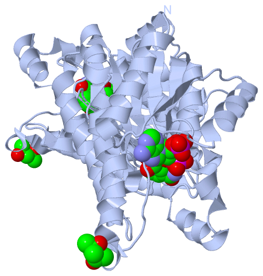
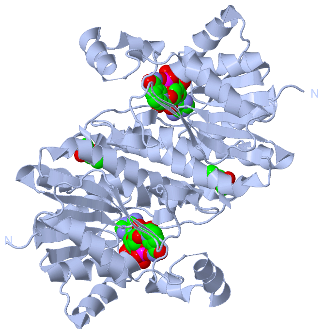
Asymmetric Unit (2, 2)
|
Sites (2, 2)
Asymmetric Unit (2, 2)
|
SS Bonds (0, 0)| (no "SS Bond" information available for 5VML) |
Cis Peptide Bonds (0, 0)| (no "Cis Peptide Bond" information available for 5VML) |
SAPs(SNPs)/Variants (0, 0)| (no "SAP(SNP)/Variant" information available for 5VML) |
PROSITE Motifs (0, 0)| (no "PROSITE Motif" information available for 5VML) |
Exons (0, 0)| (no "Exon" information available for 5VML) |
Sequences/Alignments
Asymmetric Unit
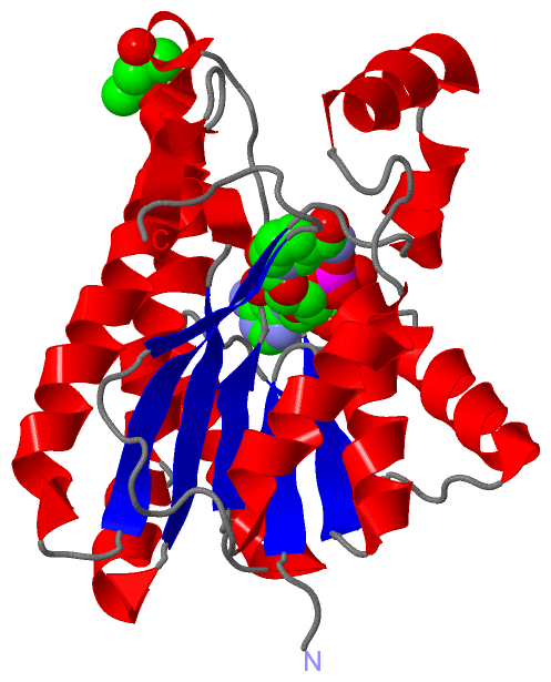
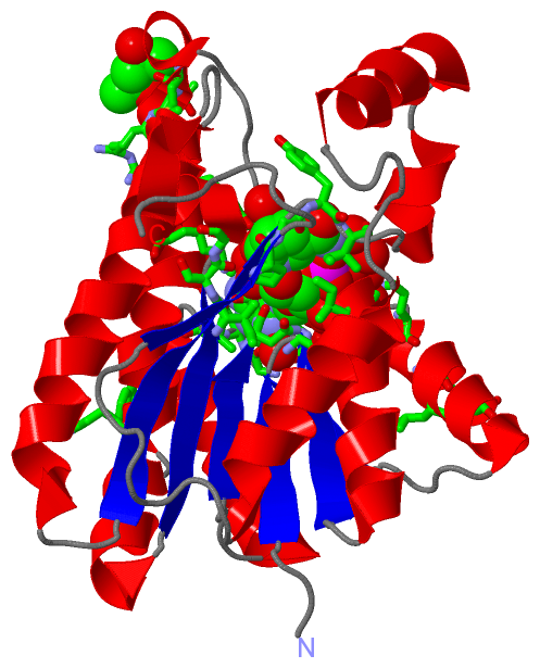
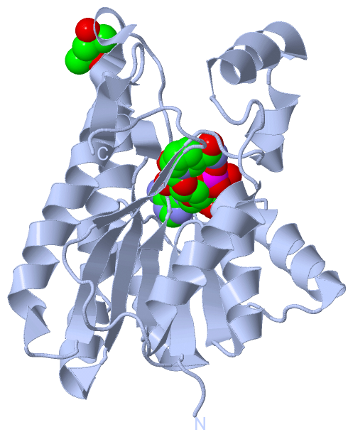
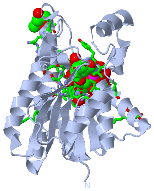
Chain A from PDB Type:PROTEIN Length:245
SCOP domains ----------------------------------------------------------------------------------------------------------------------------------------------------------------------------------------------------------------------------------------------------- SCOP domains
CATH domains ----------------------------------------------------------------------------------------------------------------------------------------------------------------------------------------------------------------------------------------------------- CATH domains
Pfam domains ----------------------------------------------------------------------------------------------------------------------------------------------------------------------------------------------------------------------------------------------------- Pfam domains
SAPs(SNPs) ----------------------------------------------------------------------------------------------------------------------------------------------------------------------------------------------------------------------------------------------------- SAPs(SNPs)
PROSITE ----------------------------------------------------------------------------------------------------------------------------------------------------------------------------------------------------------------------------------------------------- PROSITE
Transcript ----------------------------------------------------------------------------------------------------------------------------------------------------------------------------------------------------------------------------------------------------- Transcript
5vml A 4 SQRIAYVTGGMGGIGTSICQRLHKDGFRVVAGCGPNSPRRVKWLEDQKALGFDFYASEGNVGDWDSTKQAFDKVKAEVGEIDVLVNNAGITRDVVFRKMTREDWQAVIDTNLTSLFNVTKQVIDGMVERGWGRIINISSVNGQKGQFGQTNYSTAKAGIHGFTMSLAQEVATKGVTVNTVSPGYIGTDMVKAIRPDVLEKIVATIPVRRLGSPDEIGSIVAWLASEESGFSTGADFSLNGGLHMG 248
13 23 33 43 53 63 73 83 93 103 113 123 133 143 153 163 173 183 193 203 213 223 233 243
|
||||||||||||||||||||
SCOP Domains (0, 0)| (no "SCOP Domain" information available for 5VML) |
CATH Domains (0, 0)| (no "CATH Domain" information available for 5VML) |
Pfam Domains (0, 0)| (no "Pfam Domain" information available for 5VML) |
Gene Ontology (5, 5)|
Asymmetric Unit(hide GO term definitions) |
Interactive Views
|
|||||||||||||||||||||||||||||||||||||||||||||||||||||||||||||||||||||||||||||||||||||||||||||||||||||||||||||||||||||||||||||||||||||||||||||||||||||||||||||||||||||
Still Images
|
||||||||||||||||
Databases
|
||||||||||||||||||||||||||||||||||||||||||||||||||||||||||||||||||||||||||||||||||||||||||||||||||||||||||||||||||||||||||||||||||||||||||||||||||||||||||||||||
Analysis Tools
|
|||||||||||||||||||||||||||||||||||||||||||||||||||||||||||||
Entries Sharing at Least One Protein Chain (UniProt ID)
Related Entries Specified in the PDB File
|
|