Show PDB file:   
         Plain Text   HTML   (compressed file size)
QuickSearch:   
by PDB,NDB,UniProt,PROSITE Code or Search Term(s)  
(-)Asymmetric Unit
(-)Asym. Unit - sites
(-)Biological Unit 1
(-)Biol. Unit 1 - sites
(-)Biological Unit 2
collapse expand < >
Image Asymmetric Unit
Asymmetric Unit  (Jmol Viewer)
Image Asym. Unit - sites
Asym. Unit - sites  (Jmol Viewer)
Image Biological Unit 1
Biological Unit 1  (Jmol Viewer)
Image Biol. Unit 1 - sites
Biol. Unit 1 - sites  (Jmol Viewer)
Image Biological Unit 2
Biological Unit 2  (Jmol Viewer)

(-) Description

Title :  CRYSTAL STRUCTURE OF NDM-1 IN COMPLEX WITH HYDROLYZED MEROPENEM - NEW REFINEMENT
 
Authors :  J. E. Raczynska, I. G. Shabalin, M. Jaskolski, W. Minor, A. Wlodawer, D. N. C. J. Strynadka
Date :  03 Feb 17  (Deposition) - 05 Apr 17  (Release) - 05 Apr 17  (Revision)
Method :  X-RAY DIFFRACTION
Resolution :  1.90
Chains :  Asym. Unit :  A,B
Biol. Unit 1:  A  (1x)
Biol. Unit 2:  B  (1x)
Keywords :  Hydrolase-Antibiotic Complex, Metallo-Beta-Lactamase, Ndm-1 (Keyword Search: [Gene Ontology, PubMed, Web (Google))
 
Reference :  J. E. Raczynska, I. G. Shabalin, M. Jaskolski, W. Minor, A. Wlodawer
Re-Investigation Of Structural Data For Biomedically Important Enzymes - Metallo-Beta-Lactamases
To Be Published
PubMed: search

(-) Compounds

Molecule 1 - METALLO-BETA-LACTAMASE TYPE 2
    ChainsA, B
    EC Number3.5.2.6
    EngineeredYES
    Expression SystemESCHERICHIA COLI BL21
    Expression System Taxid511693
    GeneBLANDM-1
    Organism ScientificKLEBSIELLA PNEUMONIAE
    Organism Taxid573
    SynonymB2 METALLO-BETA-LACTAMASE,BETA-LACTAMASE TYPE II,METALLO- BETA-LACTAMASE NDM-1,METALLO-BETA-LACTAMASE TYPE II,NEW DELHI METALLO-BETA-LACTAMASE-1,NDM-1

 Structural Features

(-) Chains, Units

  12
Asymmetric Unit AB
Biological Unit 1 (1x)A 
Biological Unit 2 (1x) B

Summary Information (see also Sequences/Alignments below)

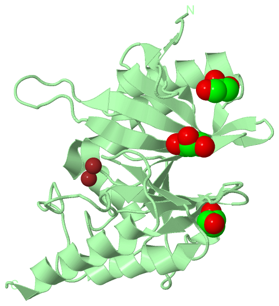
(-) Ligands, Modified Residues, Ions  (5, 12)

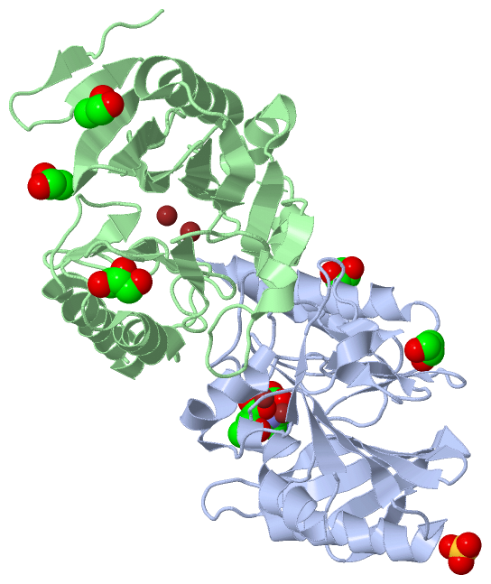
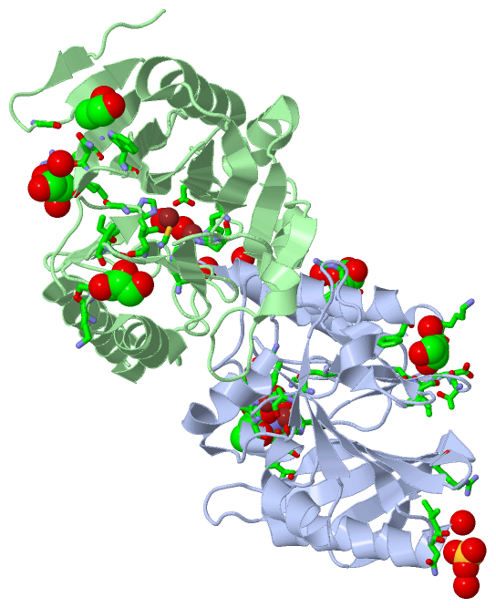
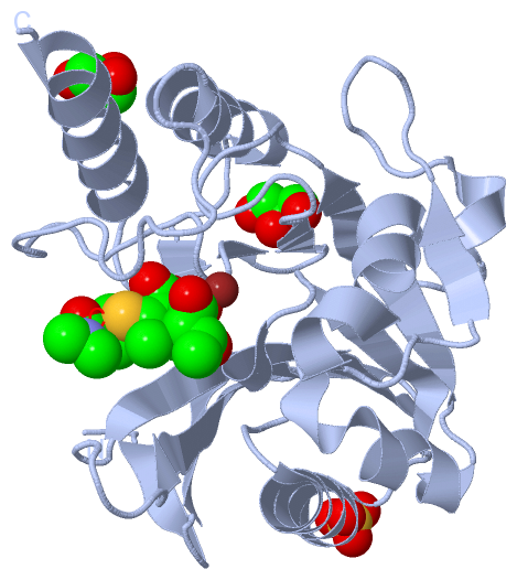
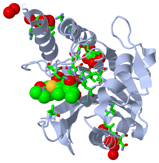
Asymmetric Unit (5, 12)
No.NameCountTypeFull Name
18FB1Ligand/Ion(3~{S},4~{R},5~{S})-5-[(2~{S},3~{R})-1,3-BIS(OXIDANYL)-1-OXIDANYLIDENE-BUTAN-2-YL]-3-[(3~{S},5~{S})-5-(DIMETHYLCARBAMOYL)PYRROLIDIN-3-YL]SULFANYL-4-METHYL-PYRROLIDINE-2-CARBOXYLIC ACID
2GOL5Ligand/IonGLYCEROL
3SO41Ligand/IonSULFATE ION
4UNK1Mod. Amino Acid
5ZN4Ligand/IonZINC ION
Biological Unit 1 (3, 4)
No.NameCountTypeFull Name
18FB1Ligand/Ion(3~{S},4~{R},5~{S})-5-[(2~{S},3~{R})-1,3-BIS(OXIDANYL)-1-OXIDANYLIDENE-BUTAN-2-YL]-3-[(3~{S},5~{S})-5-(DIMETHYLCARBAMOYL)PYRROLIDIN-3-YL]SULFANYL-4-METHYL-PYRROLIDINE-2-CARBOXYLIC ACID
2GOL2Ligand/IonGLYCEROL
3SO41Ligand/IonSULFATE ION
4UNK-1Mod. Amino Acid
5ZN-1Ligand/IonZINC ION
Biological Unit 2 (1, 3)
No.NameCountTypeFull Name
18FB-1Ligand/Ion(3~{S},4~{R},5~{S})-5-[(2~{S},3~{R})-1,3-BIS(OXIDANYL)-1-OXIDANYLIDENE-BUTAN-2-YL]-3-[(3~{S},5~{S})-5-(DIMETHYLCARBAMOYL)PYRROLIDIN-3-YL]SULFANYL-4-METHYL-PYRROLIDINE-2-CARBOXYLIC ACID
2GOL3Ligand/IonGLYCEROL
3SO4-1Ligand/IonSULFATE ION
4UNK-1Mod. Amino Acid
5ZN-1Ligand/IonZINC ION

(-) Sites  (11, 11)

Asymmetric Unit (11, 11)
No.NameEvidenceResiduesDescription
01AC1SOFTWAREVAL A:73 , HIS A:122 , ASP A:124 , HIS A:189 , CYS A:208 , LYS A:211 , ASN A:220 , HIS A:250 , ZN A:302 , ZN A:303 , HOH A:441 , HOH A:535 , HOH A:558 , HOH A:592binding site for residue 8FB A 301
02AC2SOFTWAREASP A:124 , CYS A:208 , HIS A:250 , 8FB A:301 , HOH A:535binding site for residue ZN A 302
03AC3SOFTWAREHIS A:120 , HIS A:122 , HIS A:189 , 8FB A:301 , HOH A:535binding site for residue ZN A 303
04AC4SOFTWAREARG A:234 , ALA A:263 , ARG A:264 , HOH A:406 , HOH A:423binding site for residue GOL A 304
05AC5SOFTWAREGLY A:197 , ILE A:198 , THR A:201 , ASP A:202 , ILE A:203 , PHE A:240 , LYS A:242 , ALA A:243 , HOH A:528binding site for residue GOL A 305
06AC6SOFTWAREILE A:109 , ASN A:110 , HOH A:403 , GLY B:42 , ARG B:52 , GLN B:53 , GOL B:303 , HOH B:515binding site for residue SO4 A 306
07AC7SOFTWAREASP B:124 , CYS B:208 , HIS B:250 , HOH B:534 , HOH B:542binding site for residue ZN B 301
08AC8SOFTWAREHIS B:120 , HIS B:122 , HIS B:189 , HOH B:528 , HOH B:542binding site for residue ZN B 302
09AC9SOFTWAREARG A:81 , ILE A:109 , SO4 A:306 , HOH A:471 , ARG B:52 , LEU B:54 , HOH B:405binding site for residue GOL B 303
10AD1SOFTWAREILE B:198 , THR B:201 , ILE B:203 , LYS B:242 , ALA B:243binding site for residue GOL B 304
11AD2SOFTWAREGLN B:53 , TRP B:59binding site for residue GOL B 305

(-) SS Bonds  (0, 0)

(no "SS Bond" information available for 5N0H)

(-) Cis Peptide Bonds  (0, 0)

(no "Cis Peptide Bond" information available for 5N0H)

 Sequence-Structure Mapping

(-) SAPs(SNPs)/Variants  (0, 0)

(no "SAP(SNP)/Variant" information available for 5N0H)

(-) PROSITE Motifs  (0, 0)

(no "PROSITE Motif" information available for 5N0H)

(-) Exons   (0, 0)

(no "Exon" information available for 5N0H)

(-) Sequences/Alignments

Asymmetric Unit
   Reformat: Number of residues per line =  ('0' or empty: single-line sequence representation)
  Number of residues per labelling interval =   
  UniProt sequence: complete  aligned part    
   Show mapping: SCOP domains CATH domains Pfam domains Secondary structure (by author)
SAPs(SNPs) PROSITE motifs Exons
(details for a mapped element are shown in a popup box when the mouse pointer rests over it)
Chain A from PDB  Type:PROTEIN  Length:233
                                                                                                                                                                                                                                                                         
               SCOP domains ----------------------------------------------------------------------------------------------------------------------------------------------------------------------------------------------------------------------------------------- SCOP domains
               CATH domains ----------------------------------------------------------------------------------------------------------------------------------------------------------------------------------------------------------------------------------------- CATH domains
               Pfam domains ----------------------------------------------------------------------------------------------------------------------------------------------------------------------------------------------------------------------------------------- Pfam domains
         Sec.struct. author ..eeee..eeeeeee..eeeeeeee.......eeeeeeeeee..eeeee....hhhhhhhhhhhhhhhh...eeeeee...hhhhhhhhhhhhhh..eeeeehhhhhhhhhhh.....eee........hhhhh.....eeee...........eeee....eee......................hhhhhhhhhhhhh....eee........hhhhhhhhhhhhhhhhh. Sec.struct. author
                 SAPs(SNPs) ----------------------------------------------------------------------------------------------------------------------------------------------------------------------------------------------------------------------------------------- SAPs(SNPs)
                    PROSITE ----------------------------------------------------------------------------------------------------------------------------------------------------------------------------------------------------------------------------------------- PROSITE
                 Transcript ----------------------------------------------------------------------------------------------------------------------------------------------------------------------------------------------------------------------------------------- Transcript
                 5n0h A  41 TGDQRFGDLVFRQLAPNVWQHTSYLDMPGFGAVASNGLIVRDGGRVLVVDTAWTDDQTAQILNWIKQEINLPVALAVVTHAHQDKMGGMDALHAAGIATYANALSNQLAPQEGMVAAQHSLTFAANGWVEPATAPNFGPLKVFYPGPGHTSDNITVGIDGTDIAFGGCLIKDSKAKSLGNLGDADTEHYAASARAFGAAFPKASMIVMSHSAPDSRAAITHTARMADKLRKLx 273
                                    50        60        70        80        90       100       110       120       130       140       150       160       170       180       190       200       210       220       230       240       250       260       270  |
                                                                                                                                                                                                                                                                  273-UNK

Chain B from PDB  Type:PROTEIN  Length:240
                                                                                                                                                                                                                                                                                
               SCOP domains ------------------------------------------------------------------------------------------------------------------------------------------------------------------------------------------------------------------------------------------------ SCOP domains
               CATH domains ------------------------------------------------------------------------------------------------------------------------------------------------------------------------------------------------------------------------------------------------ CATH domains
               Pfam domains ------------------------------------------------------------------------------------------------------------------------------------------------------------------------------------------------------------------------------------------------ Pfam domains
         Sec.struct. author ....ee.....eee..eeeeeee..eeeeeee.........eeeeeeeee..eeeee....hhhhhhhhhhhhhhhh...eeeee....hhhhhhhhhhhhhh..eeeeehhhhhhhhhhh.....eee........hhhhh.....eeee...........eeee....eeeee....................hhhhhhhhhhhhh....eeee.......hhhhhhhhhhhhhhhh. Sec.struct. author
                 SAPs(SNPs) ------------------------------------------------------------------------------------------------------------------------------------------------------------------------------------------------------------------------------------------------ SAPs(SNPs)
                    PROSITE ------------------------------------------------------------------------------------------------------------------------------------------------------------------------------------------------------------------------------------------------ PROSITE
                 Transcript ------------------------------------------------------------------------------------------------------------------------------------------------------------------------------------------------------------------------------------------------ Transcript
                 5n0h B  33 PTIGQQMETGDQRFGDLVFRQLAPNVWQHTSYLDMPGFGAVASNGLIVRDGGRVLVVDTAWTDDQTAQILNWIKQEINLPVALAVVTHAHQDKMGGMDALHAAGIATYANALSNQLAPQEGMVAAQHSLTFAANGWVEPATAPNFGPLKVFYPGPGHTSDNITVGIDGTDIAFGGCLIKDSKAKSLGNLGDADTEHYAASARAFGAAFPKASMIVMSHSAPDSRAAITHTARMADKLRKL 272
                                    42        52        62        72        82        92       102       112       122       132       142       152       162       172       182       192       202       212       222       232       242       252       262       272

   Legend:   → Mismatch (orange background)
  - → Gap (green background, '-', border residues have a numbering label)
    → Modified Residue (blue background, lower-case, 'x' indicates undefined single-letter code, labelled with number + name)
  x → Chemical Group (purple background, 'x', labelled with number + name, e.g. ACE or NH2)
  extra numbering lines below/above indicate numbering irregularities and modified residue names etc., number ends below/above '|'

 Classification and Annotation

(-) SCOP Domains  (0, 0)

(no "SCOP Domain" information available for 5N0H)

(-) CATH Domains  (0, 0)

(no "CATH Domain" information available for 5N0H)

(-) Pfam Domains  (0, 0)

(no "Pfam Domain" information available for 5N0H)

(-) Gene Ontology  (7, 7)

Asymmetric Unit(hide GO term definitions)

 Visualization

(-) Interactive Views

Asymmetric Unit
  Complete Structure
    Jena3D(integrated viewing of ligand, site, SAP, PROSITE, SCOP information)
    WebMol | AstexViewer[tm]@PDBe
(Java Applets, require no local installation except for Java; loading may be slow)
    STRAP
(Java WebStart application, automatic local installation, requires Java; full application with system access!)
    RasMol
(require local installation)
    Molscript (VRML)
(requires installation of a VRML viewer; select preferred view via VRML and generate a mono or stereo PDF format file)
 
  Ligands, Modified Residues, Ions
    8FB  [ RasMol | Jena3D ]  +environment [ RasMol | Jena3D ]
    GOL  [ RasMol | Jena3D ]  +environment [ RasMol | Jena3D ]
    SO4  [ RasMol | Jena3D ]  +environment [ RasMol | Jena3D ]
    UNK  [ RasMol | Jena3D ]  +environment [ RasMol | Jena3D ]
    ZN  [ RasMol | Jena3D ]  +environment [ RasMol | Jena3D ]
 
  Sites
    AC1  [ RasMol ]  +environment [ RasMol ]
    AC2  [ RasMol ]  +environment [ RasMol ]
    AC3  [ RasMol ]  +environment [ RasMol ]
    AC4  [ RasMol ]  +environment [ RasMol ]
    AC5  [ RasMol ]  +environment [ RasMol ]
    AC6  [ RasMol ]  +environment [ RasMol ]
    AC7  [ RasMol ]  +environment [ RasMol ]
    AC8  [ RasMol ]  +environment [ RasMol ]
    AC9  [ RasMol ]  +environment [ RasMol ]
    AD1  [ RasMol ]  +environment [ RasMol ]
    AD2  [ RasMol ]  +environment [ RasMol ]
 
  Cis Peptide Bonds
(no "Cis Peptide Bonds" information available for 5n0h)
 
Biological Units
  Complete Structure
    Biological Unit 1  [ Jena3D ]
    Biological Unit 2  [ Jena3D ]

(-) Still Images

Jmol
  protein: cartoon or spacefill or dots and stick; nucleic acid: cartoon and stick; ligands: spacefill; active site: stick
Molscript
  protein, nucleic acid: cartoon; ligands: spacefill; active site: ball and stick

 Databases and Analysis Tools

(-) Databases

Access by PDB/NDB ID
  5n0h
    Family and Domain InformationProDom | SYSTERS
    General Structural InformationGlycoscienceDB | MMDB | NDB | OCA | PDB | PDBe | PDBj | PDBsum | PDBWiki | PQS | PROTEOPEDIA
    Orientation in MembranesOPM
    Protein SurfaceSURFACE
    Secondary StructureDSSP (structure derived) | HSSP (homology derived)
    Structural GenomicsGeneCensus
    Structural NeighboursCE | VAST
    Structure ClassificationCATH | Dali | SCOP
    Validation and Original DataBMRB Data View | BMRB Restraints Grid | EDS | PROCHECK | RECOORD | WHAT_CHECK
 
Access by UniProt ID/Accession number
  BLAN1_KLEPN | C7C422
    Comparative Protein Structure ModelsModBase
    Genomic InformationEnsembl
    Protein-protein InteractionDIP
    Sequence, Family and Domain InformationInterPro | Pfam | SMART | UniProtKB/SwissProt
 
Access by Enzyme Classificator   (EC Number)
  3.5.2.6
    General Enzyme InformationBRENDA | EC-PDB | Enzyme | IntEnz
    PathwayKEGG | MetaCyc
 
Access by Disease Identifier   (MIM ID)
  (no 'MIM ID' available)
    Disease InformationOMIM
 
Access by GenAge ID
  (no 'GenAge ID' available)
    Age Related InformationGenAge

(-) Analysis Tools

Access by PDB/NDB ID
    Domain InformationXDom
    Interatomic Contacts of Structural UnitsCSU
    Ligand-protein ContactsLPC
    Protein CavitiescastP
    Sequence and Secondary StructurePDBCartoon
    Structure AlignmentSTRAP(Java WebStart application, automatic local installation, requires Java; full application with system access!)
    Structure and Sequence BrowserSTING
 
Access by UniProt ID/Accession number
  BLAN1_KLEPN | C7C422
    Protein Disorder PredictionDisEMBL | FoldIndex | GLOBPLOT (for more information see DisProt)

 Related Entries

(-) Entries Sharing at Least One Protein Chain (UniProt ID)

UniProtKB/Swiss-Prot
        BLAN1_KLEPN | C7C4223pg4 3q6x 3rkj 3rkk 3sbl 3sfp 3spu 3srx 3zr9 4exs 4exy 4ey2 4eyb 4eyf 4eyl 4gyq 4gyu 4h0d 4hky 4hl1 4hl2 4ram 4raw 4rbs 4rl0 4rl2 4rm5 4u4l 5a5z 5jqj 5k4k 5k4m 5k4n 5n0i

(-) Related Entries Specified in the PDB File

4eyl