|
|
|
|
 Description
|
|
Compounds
|
||||||||||||||||||||||||||||||||||||||||||||||||
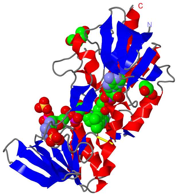
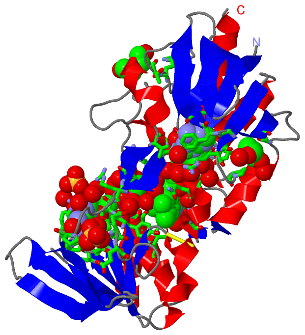
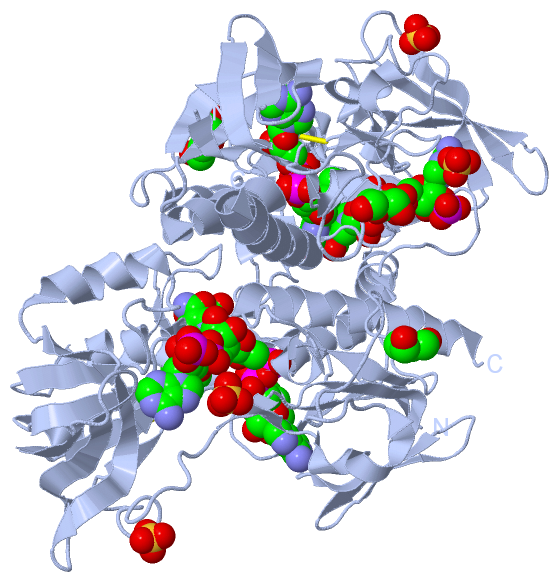
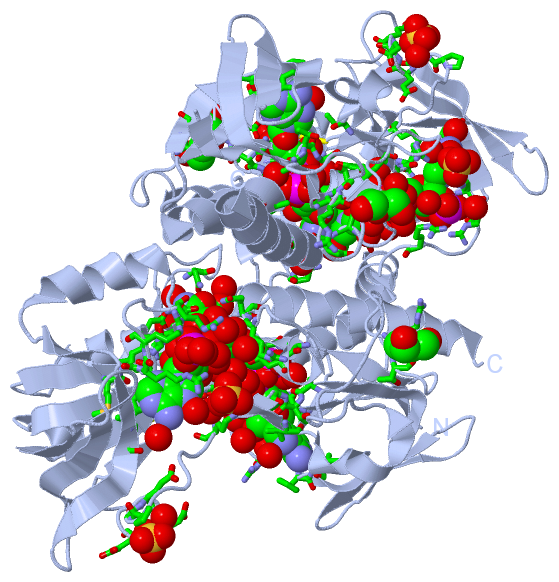
Chains, Units
Summary Information (see also Sequences/Alignments below) |
Ligands, Modified Residues, Ions (4, 6)| Asymmetric Unit (4, 6) Biological Unit 1 (4, 12) |
Sites (6, 6)
Asymmetric Unit (6, 6)
|
SS Bonds (1, 1)
Asymmetric Unit
|
||||||||
Cis Peptide Bonds (0, 0)| (no "Cis Peptide Bond" information available for 5MIQ) |
SAPs(SNPs)/Variants (0, 0)| (no "SAP(SNP)/Variant" information available for 5MIQ) |
PROSITE Motifs (0, 0)| (no "PROSITE Motif" information available for 5MIQ) |
Exons (0, 0)| (no "Exon" information available for 5MIQ) |
Sequences/Alignments
Asymmetric Unit
Chain A from PDB Type:PROTEIN Length:303
SCOP domains --------------------------------------------------------------------------------------------------------------------------------------------------------------------------------------------------------------------------------------------------------------------------------------------------------------- SCOP domains
CATH domains --------------------------------------------------------------------------------------------------------------------------------------------------------------------------------------------------------------------------------------------------------------------------------------------------------------- CATH domains
Pfam domains --------------------------------------------------------------------------------------------------------------------------------------------------------------------------------------------------------------------------------------------------------------------------------------------------------------- Pfam domains
SAPs(SNPs) --------------------------------------------------------------------------------------------------------------------------------------------------------------------------------------------------------------------------------------------------------------------------------------------------------------- SAPs(SNPs)
PROSITE --------------------------------------------------------------------------------------------------------------------------------------------------------------------------------------------------------------------------------------------------------------------------------------------------------------- PROSITE
Transcript --------------------------------------------------------------------------------------------------------------------------------------------------------------------------------------------------------------------------------------------------------------------------------------------------------------- Transcript
5miq A 5 KYDVVIIGSGPAGMTAAMYTARSEMKTLLLERGVPGGQMNNTAEIENYPGYETIMGPELSMKMAEPLEGLGVENAYGFVTGIEDHGDYKKIITEDDEFITKSIIIATGANHRKLEIPGEEEYGARGVSYCAVCDGAFFRNQEILVIGGGDSAVEEALYLTRFGQSVTIMHRRDKLRAQEIIQQRAFKEEKINFIWDSVPMEIKGDDKKIQSVVYKNVKTGEVTEKAFGGIFIYVGLDPVAEFVSDLGITDEAGWIITDDHMRTNIPGIFAVGDVRQKDFRQITTAVGDGAQAAQEAYKFVVEL 307
14 24 34 44 54 64 74 84 94 104 114 124 134 144 154 164 174 184 194 204 214 224 234 244 254 264 274 284 294 304
|
||||||||||||||||||||
SCOP Domains (0, 0)| (no "SCOP Domain" information available for 5MIQ) |
CATH Domains (0, 0)| (no "CATH Domain" information available for 5MIQ) |
Pfam Domains (0, 0)| (no "Pfam Domain" information available for 5MIQ) |
Gene Ontology (0, 0)|
Asymmetric Unit(hide GO term definitions)
(no "Gene Ontology" information available for 5MIQ)
|
Interactive Views
|
||||||||||||||||||||||||||||||||||||||||||||||||||||||||||||||||||||||||||||||||||||||||||||||||||||||||||||||||||||||||||||||||||||||||||||||||||||||||||||||||||||||||||||||||||||||||||||||||
Still Images
|
||||||||||||||||
Databases
|
||||||||||||||||||||||||||||||||||||||||||||||||||||||||||||||||||||||||||||||||||||||||||||||||||||||||||||||||||||||||||||||||||||||||||||||||||||||||||||||||
Analysis Tools
|
|||||||||||||||||||||||||||||||||||||||||||||||||||||||||||||
Entries Sharing at Least One Protein Chain (UniProt ID)
Related Entries Specified in the PDB File
|
|