|
|
|
|
 Description
|
|
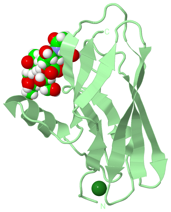
Compounds
|
||||||||||||||||||||||||||||||||||||||||||||||||||||
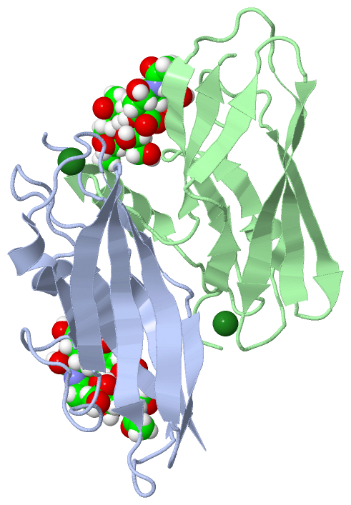
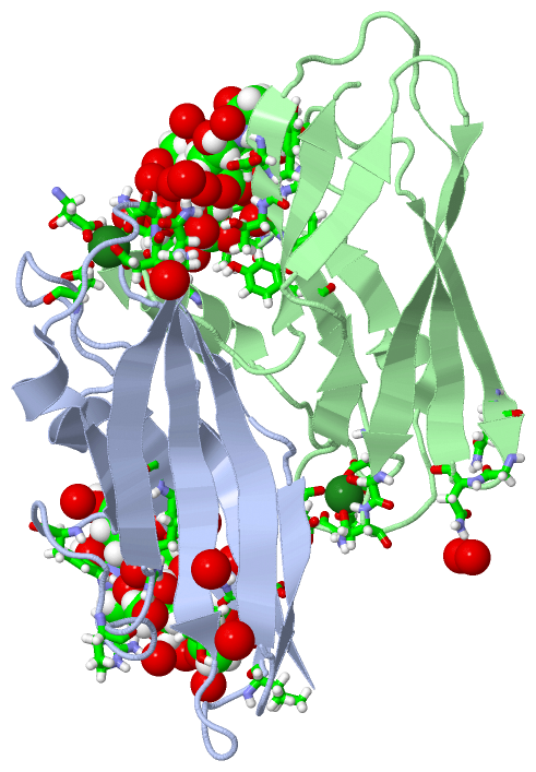
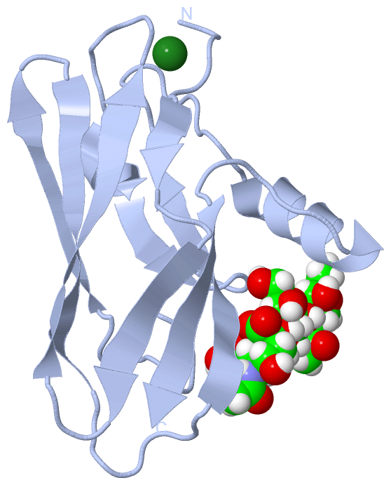
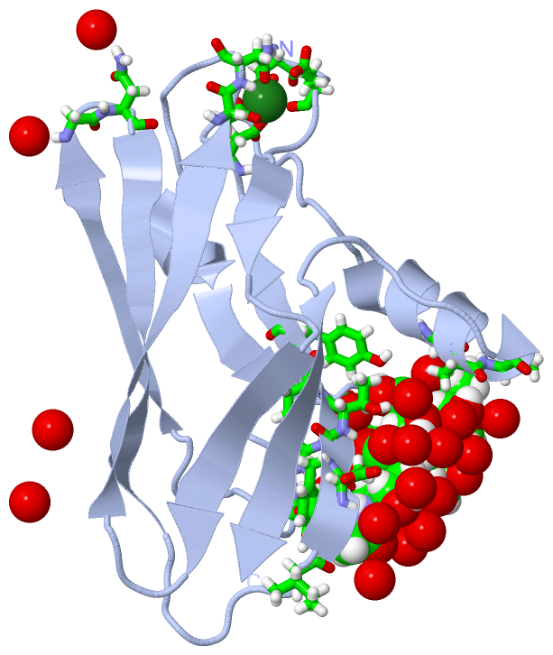
Chains, Units
Summary Information (see also Sequences/Alignments below) |
Ligands, Modified Residues, Ions (4, 8)
Asymmetric Unit (4, 8)
|
Sites (4, 4)
Asymmetric Unit (4, 4)
|
SS Bonds (0, 0)| (no "SS Bond" information available for 5IUC) |
Cis Peptide Bonds (0, 0)| (no "Cis Peptide Bond" information available for 5IUC) |
SAPs(SNPs)/Variants (0, 0)| (no "SAP(SNP)/Variant" information available for 5IUC) |
PROSITE Motifs (0, 0)| (no "PROSITE Motif" information available for 5IUC) |
Exons (0, 0)| (no "Exon" information available for 5IUC) |
Sequences/Alignments
Asymmetric Unit
Chain A from PDB Type:PROTEIN Length:123
SCOP domains --------------------------------------------------------------------------------------------------------------------------- SCOP domains
CATH domains --------------------------------------------------------------------------------------------------------------------------- CATH domains
Pfam domains --------------------------------------------------------------------------------------------------------------------------- Pfam domains
SAPs(SNPs) --------------------------------------------------------------------------------------------------------------------------- SAPs(SNPs)
PROSITE --------------------------------------------------------------------------------------------------------------------------- PROSITE
Transcript --------------------------------------------------------------------------------------------------------------------------- Transcript
5iuc A 399 DTERPVVNVPSEITVYRGESFEYFATVTDNSNAFDLAKTVVRWLYSNQPGRGTEWLQYSVTQVGNQLKVRIFGNVPIDTTIGDYTRYVVATDAAGNVNATQTEMGNAAVDKTSVNGQFKLIIR 521
408 418 428 438 448 458 468 478 488 498 508 518
Chain B from PDB Type:PROTEIN Length:123
SCOP domains --------------------------------------------------------------------------------------------------------------------------- SCOP domains
CATH domains --------------------------------------------------------------------------------------------------------------------------- CATH domains
Pfam domains --------------------------------------------------------------------------------------------------------------------------- Pfam domains
SAPs(SNPs) --------------------------------------------------------------------------------------------------------------------------- SAPs(SNPs)
PROSITE --------------------------------------------------------------------------------------------------------------------------- PROSITE
Transcript --------------------------------------------------------------------------------------------------------------------------- Transcript
5iuc B 399 DTERPVVNVPSEITVYRGESFEYFATVTDNSNAFDLAKTVVRWLYSNQPGRGTEWLQYSVTQVGNQLKVRIFGNVPIDTTIGDYTRYVVATDAAGNVNATQTEMGNAAVDKTSVNGQFKLIIR 521
408 418 428 438 448 458 468 478 488 498 508 518
|
||||||||||||||||||||
SCOP Domains (0, 0)| (no "SCOP Domain" information available for 5IUC) |
CATH Domains (0, 0)| (no "CATH Domain" information available for 5IUC) |
Pfam Domains (0, 0)| (no "Pfam Domain" information available for 5IUC) |
Gene Ontology (3, 3)|
Asymmetric Unit(hide GO term definitions) |
Interactive Views
|
|||||||||||||||||||||||||||||||||||||||||||||||||||||||||||||||||||||||||||||||||||||||||||||||||||||||||||||||||||||||||||||||||||||||||||||||||||||||||||||||||||||||||||||||||||||||
Still Images
|
||||||||||||||||
Databases
|
||||||||||||||||||||||||||||||||||||||||||||||||||||||||||||||||||||||||||||||||||||||||||||||||||||||||||||||||||||||||||||||||||||||||||||||||||||||||||||||||
Analysis Tools
|
|||||||||||||||||||||||||||||||||||||||||||||||||||||||||||||
Entries Sharing at Least One Protein Chain (UniProt ID)
Related Entries Specified in the PDB File
|
|