|
|
|
|
 Description
|
|
Compounds
|
||||||||||||||||||||||||||||||||||||||||
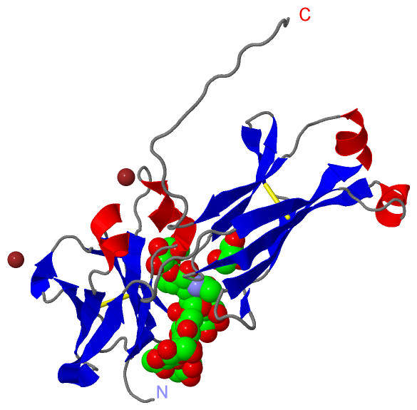
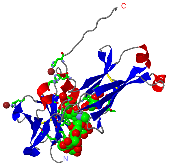
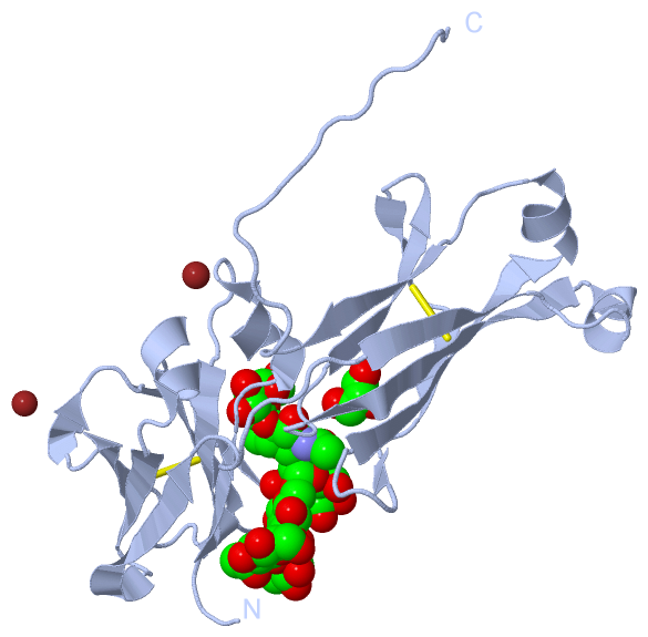
Chains, Units
Summary Information (see also Sequences/Alignments below) |
Ligands, Modified Residues, Ions (7, 12)
Asymmetric Unit (7, 12)
|
Sites (4, 4)
Asymmetric Unit (4, 4)
|
SS Bonds (2, 2)
Asymmetric Unit
|
||||||||||||
Cis Peptide Bonds (1, 1)
Asymmetric Unit
|
||||||||
SAPs(SNPs)/Variants (0, 0)| (no "SAP(SNP)/Variant" information available for 5HVW) |
PROSITE Motifs (0, 0)| (no "PROSITE Motif" information available for 5HVW) |
Exons (0, 0)| (no "Exon" information available for 5HVW) |
Sequences/Alignments
Asymmetric Unit
Chain A from PDB Type:PROTEIN Length:210
SCOP domains ------------------------------------------------------------------------------------------------------------------------------------------------------------------------------------------------------------------ SCOP domains
CATH domains ------------------------------------------------------------------------------------------------------------------------------------------------------------------------------------------------------------------ CATH domains
Pfam domains ------------------------------------------------------------------------------------------------------------------------------------------------------------------------------------------------------------------ Pfam domains
SAPs(SNPs) ------------------------------------------------------------------------------------------------------------------------------------------------------------------------------------------------------------------ SAPs(SNPs)
PROSITE ------------------------------------------------------------------------------------------------------------------------------------------------------------------------------------------------------------------ PROSITE
Transcript ------------------------------------------------------------------------------------------------------------------------------------------------------------------------------------------------------------------ Transcript
5hvw A 235 LGGPSVFLFPPKPKDTLMISRTPEVTCVVVDVSQEDPEVQFNWYVDGVEVHNAKTKPREEQFNSTYRVVSVLTVLHQDWLNGKEYKCKVSNKGLPSSIEKTISKAKGQPREPQVYTFPPSQEEMTKNQVSLRCLVKGFYPSDIAVEWESNGQPENNYKTTKPVLDSDGSFRLESRLTVDKSRWQEGNVFSCSVMHEALHNHYTQKSLSLS 444
244 254 264 274 284 294 304 314 324 334 344 354 364 374 384 394 404 414 424 434 444
|
||||||||||||||||||||
SCOP Domains (0, 0)| (no "SCOP Domain" information available for 5HVW) |
CATH Domains (0, 0)| (no "CATH Domain" information available for 5HVW) |
Pfam Domains (0, 0)| (no "Pfam Domain" information available for 5HVW) |
Gene Ontology (19, 19)|
Asymmetric Unit(hide GO term definitions) |
Interactive Views
|
|||||||||||||||||||||||||||||||||||||||||||||||||||||||||||||||||||||||||||||||||||||||||||||||||||||||||||||||||||||||||||||||||||||||||||||||||||||||||||||||||||||||||||||||||||||||||||||||||||||||||||||
Still Images
|
||||||||||||||||
Databases
|
||||||||||||||||||||||||||||||||||||||||||||||||||||||||||||||||||||||||||||||||||||||||||||||||||||||||||||||||||||||||||||||||||||||||||||||||||||||||||||||||
Analysis Tools
|
|||||||||||||||||||||||||||||||||||||||||||||||||||||||||||||
Entries Sharing at Least One Protein Chain (UniProt ID)
Related Entries Specified in the PDB File
|
|