Show PDB file:   
         Plain Text   HTML   (compressed file size)
QuickSearch:   
by PDB,NDB,UniProt,PROSITE Code or Search Term(s)  
(-)Asymmetric Unit
(-)Asym. Unit - sites
(-)Biological Unit 1
(-)Biol. Unit 1 - sites
(-)Biological Unit 2
collapse expand < >
Image Asymmetric Unit
Asymmetric Unit  (Jmol Viewer)
Image Asym. Unit - sites
Asym. Unit - sites  (Jmol Viewer)
Image Biological Unit 1
Biological Unit 1  (Jmol Viewer)
Image Biol. Unit 1 - sites
Biol. Unit 1 - sites  (Jmol Viewer)
Image Biological Unit 2
Biological Unit 2  (Jmol Viewer)

(-) Description

Title :  CRYSTAL STRUCTURE OF N-TERMINAL DOMAIN OF HUMAN PROTEIN O-MANNOSE BETA-1,2-N-ACETYLGLUCOSAMINYLTRANSFERASE IN COMPLEX WITH MAN-BETA-PNP
 
Authors :  N. Kuwabara, T. Senda, R. Kato
Date :  16 Jun 16  (Deposition) - 10 Aug 16  (Release) - 31 Aug 16  (Revision)
Method :  X-RAY DIFFRACTION
Resolution :  1.30
Chains :  Asym. Unit :  A,B
Biol. Unit 1:  A  (1x)
Biol. Unit 2:  B  (1x)
Keywords :  Glycosyltransferease, O-Mannosylation, Alpha-Dystroglycan, Sugar Binding Protein (Keyword Search: [Gene Ontology, PubMed, Web (Google))
 
Reference :  N. Kuwabara, H. Manya, T. Yamada, H. Tateno, M. Kanagawa, K. Kobayashi K. Akasaka-Manya, Y. Hirose, M. Mizuno, M. Ikeguchi, T. Toda, J. Hirabayashi, T. Senda, T. Endo, R. Kato
Carbohydrate-Binding Domain Of The Pomgnt1 Stem Region Modulates O-Mannosylation Sites Of Alpha-Dystroglycan
Proc. Natl. Acad. Sci. Usa V. 113 9280 2016
PubMed-ID: 27493216  |  Reference-DOI: 10.1073/PNAS.1525545113

(-) Compounds

Molecule 1 - PROTEIN O-LINKED-MANNOSE BETA-1,2-N- ACETYLGLUCOSAMINYLTRANSFERASE 1
    ChainsA, B
    EC Number2.4.1.-
    EngineeredYES
    Expression SystemESCHERICHIA COLI
    Expression System Taxid562
    Expression System Vector TypePLASMID
    FragmentUNP RESIDUES 92-250
    GenePOMGNT1, MGAT1.2, UNQ746/PRO1475
    Organism CommonHUMAN
    Organism ScientificHOMO SAPIENS
    Organism Taxid9606

 Structural Features

(-) Chains, Units

  12
Asymmetric Unit AB
Biological Unit 1 (1x)A 
Biological Unit 2 (1x) B

Summary Information (see also Sequences/Alignments below)

(-) Ligands, Modified Residues, Ions  (4, 8)

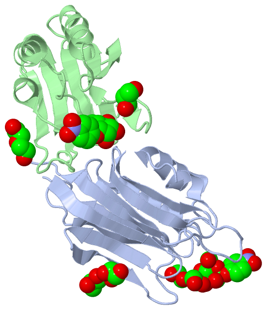
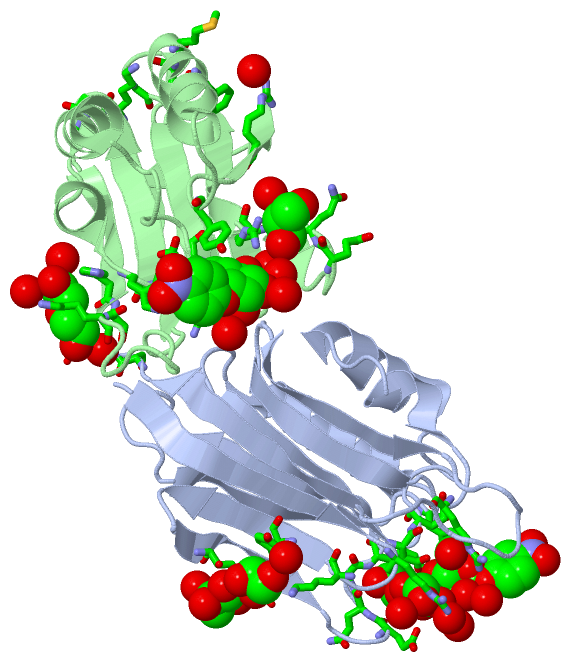
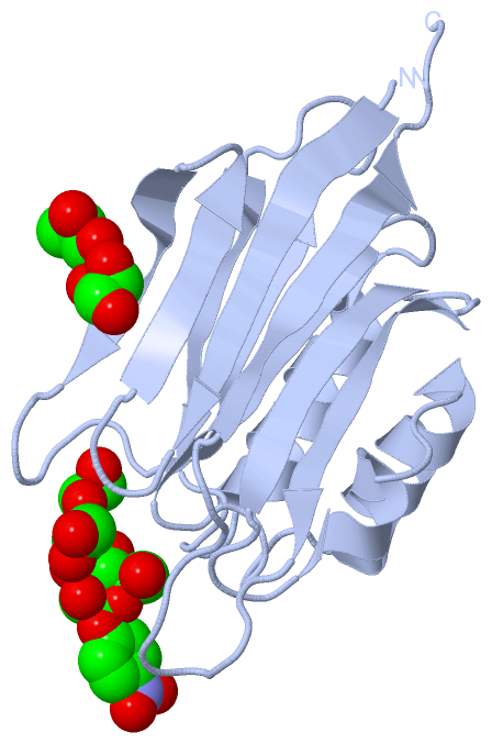
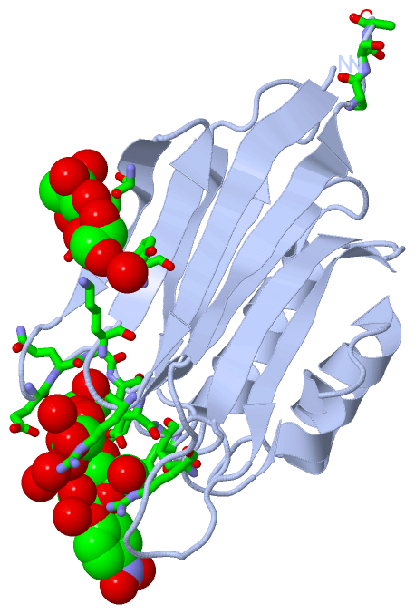
Asymmetric Unit (4, 8)
No.NameCountTypeFull Name
1EDO2Ligand/Ion1,2-ETHANEDIOL
2GOL3Ligand/IonGLYCEROL
3MBE2Ligand/Ion4-NITROPHENYL-BETA-D-MANNOPYRANOSIDE
4PEG1Ligand/IonDI(HYDROXYETHYL)ETHER
Biological Unit 1 (3, 5)
No.NameCountTypeFull Name
1EDO2Ligand/Ion1,2-ETHANEDIOL
2GOL2Ligand/IonGLYCEROL
3MBE1Ligand/Ion4-NITROPHENYL-BETA-D-MANNOPYRANOSIDE
4PEG-1Ligand/IonDI(HYDROXYETHYL)ETHER
Biological Unit 2 (3, 3)
No.NameCountTypeFull Name
1EDO-1Ligand/Ion1,2-ETHANEDIOL
2GOL1Ligand/IonGLYCEROL
3MBE1Ligand/Ion4-NITROPHENYL-BETA-D-MANNOPYRANOSIDE
4PEG1Ligand/IonDI(HYDROXYETHYL)ETHER

(-) Sites  (8, 8)

Asymmetric Unit (8, 8)
No.NameEvidenceResiduesDescription
1AC1SOFTWAREARG A:129 , TYR A:152 , ASP A:179 , GLU A:180 , ARG A:207 , EDO A:304 , HOH A:457 , HOH A:459 , HOH A:508 , GLY B:170 , ARG B:215 , LYS B:216 , GLY B:217 , GLY B:218binding site for residue MBE A 301
2AC2SOFTWARELYS A:109 , TYR A:111 , GLU A:121 , GOL A:303 , HOH A:492 , ARG B:147 , MET B:166 , HOH B:503binding site for residue GOL A 302
3AC3SOFTWARETHR A:118 , GLU A:121 , GOL A:302 , HOH A:405 , HOH A:417 , PHE B:163 , MET B:166 , VAL B:167binding site for residue GOL A 303
4AC4SOFTWAREARG A:107 , ASP A:179 , MBE A:301 , HOH A:424 , HOH A:427binding site for residue EDO A 304
5AC5SOFTWAREGLU A:126 , GLN A:127 , GLY A:128 , ARG A:129 , HOH A:447binding site for residue EDO A 305
6AC6SOFTWARESER A:247 , SER A:248 , ALA A:249 , ARG B:129 , TYR B:152 , ASP B:179 , GLU B:180 , TRP B:206 , ARG B:207 , HOH B:417 , HOH B:446 , HOH B:497binding site for residue MBE B 301
7AC7SOFTWAREGLU B:126 , GLN B:127 , GLY B:128 , ARG B:129 , HOH B:434 , HOH B:441 , HOH B:446binding site for residue GOL B 302
8AC8SOFTWAREHIS B:225 , SER B:226 , LYS B:227 , HOH B:442 , HOH B:488 , HOH B:495 , HOH B:498binding site for residue PEG B 303

(-) SS Bonds  (0, 0)

(no "SS Bond" information available for 5GGK)

(-) Cis Peptide Bonds  (2, 2)

Asymmetric Unit
No.Residues
1Gly A:218 -Pro A:219
2Gly B:218 -Pro B:219

 Sequence-Structure Mapping

(-) SAPs(SNPs)/Variants  (0, 0)

(no "SAP(SNP)/Variant" information available for 5GGK)

(-) PROSITE Motifs  (0, 0)

(no "PROSITE Motif" information available for 5GGK)

(-) Exons   (0, 0)

(no "Exon" information available for 5GGK)

(-) Sequences/Alignments

Asymmetric Unit
   Reformat: Number of residues per line =  ('0' or empty: single-line sequence representation)
  Number of residues per labelling interval =   
  UniProt sequence: complete  aligned part    
   Show mapping: SCOP domains CATH domains Pfam domains Secondary structure (by author)
SAPs(SNPs) PROSITE motifs Exons
(details for a mapped element are shown in a popup box when the mouse pointer rests over it)
Chain A from PDB  Type:PROTEIN  Length:153
                                                                                                                                                                                         
               SCOP domains --------------------------------------------------------------------------------------------------------------------------------------------------------- SCOP domains
               CATH domains --------------------------------------------------------------------------------------------------------------------------------------------------------- CATH domains
               Pfam domains --------------------------------------------------------------------------------------------------------------------------------------------------------- Pfam domains
         Sec.struct. author .eeeeeeee....eeeee..eeeeee.......eeeeeee......eeeeeee.....hhhhhhhhhhhhh....eeeeeee.......hhhhhhhhhhh...hhhhh....eeeeeee....eeeeeee...........eeeeeeee.... Sec.struct. author
                 SAPs(SNPs) --------------------------------------------------------------------------------------------------------------------------------------------------------- SAPs(SNPs)
                    PROSITE --------------------------------------------------------------------------------------------------------------------------------------------------------- PROSITE
                 Transcript --------------------------------------------------------------------------------------------------------------------------------------------------------- Transcript
                 5ggk A  97 RVLDVEVYSSRSKVYVAVDGTTVLEDEAREQGRGIHVIVLNQATGHVMAKRVFDTYSPHEDEAMVLFLNMVAPGRVLICTVKDEGSFHLKDTAKALLRSLGSQAGPALGWRDTWAFVGRKGGPVFGEKHSKSPALSSWGDPVLLKTDVPLSSA 249
                                   106       116       126       136       146       156       166       176       186       196       206       216       226       236       246   

Chain B from PDB  Type:PROTEIN  Length:152
                                                                                                                                                                                        
               SCOP domains -------------------------------------------------------------------------------------------------------------------------------------------------------- SCOP domains
               CATH domains -------------------------------------------------------------------------------------------------------------------------------------------------------- CATH domains
               Pfam domains -------------------------------------------------------------------------------------------------------------------------------------------------------- Pfam domains
         Sec.struct. author .eeeeeeee....eeeee..eeeeee.......eeeeeee......eeeeeee.....hhhhhhhhhhhhh....eeeeeee.......hhhhhhhhhhh...hhhhh....eeeeeee....eeeeeee...........eeeeeeee... Sec.struct. author
                 SAPs(SNPs) -------------------------------------------------------------------------------------------------------------------------------------------------------- SAPs(SNPs)
                    PROSITE -------------------------------------------------------------------------------------------------------------------------------------------------------- PROSITE
                 Transcript -------------------------------------------------------------------------------------------------------------------------------------------------------- Transcript
                 5ggk B  97 RVLDVEVYSSRSKVYVAVDGTTVLEDEAREQGRGIHVIVLNQATGHVMAKRVFDTYSPHEDEAMVLFLNMVAPGRVLICTVKDEGSFHLKDTAKALLRSLGSQAGPALGWRDTWAFVGRKGGPVFGEKHSKSPALSSWGDPVLLKTDVPLSS 248
                                   106       116       126       136       146       156       166       176       186       196       206       216       226       236       246  

   Legend:   → Mismatch (orange background)
  - → Gap (green background, '-', border residues have a numbering label)
    → Modified Residue (blue background, lower-case, 'x' indicates undefined single-letter code, labelled with number + name)
  x → Chemical Group (purple background, 'x', labelled with number + name, e.g. ACE or NH2)
  extra numbering lines below/above indicate numbering irregularities and modified residue names etc., number ends below/above '|'

 Classification and Annotation

(-) SCOP Domains  (0, 0)

(no "SCOP Domain" information available for 5GGK)

(-) CATH Domains  (0, 0)

(no "CATH Domain" information available for 5GGK)

(-) Pfam Domains  (0, 0)

(no "Pfam Domain" information available for 5GGK)

(-) Gene Ontology  (11, 11)

Asymmetric Unit(hide GO term definitions)

 Visualization

(-) Interactive Views

Asymmetric Unit
  Complete Structure
    Jena3D(integrated viewing of ligand, site, SAP, PROSITE, SCOP information)
    WebMol | AstexViewer[tm]@PDBe
(Java Applets, require no local installation except for Java; loading may be slow)
    STRAP
(Java WebStart application, automatic local installation, requires Java; full application with system access!)
    RasMol
(require local installation)
    Molscript (VRML)
(requires installation of a VRML viewer; select preferred view via VRML and generate a mono or stereo PDF format file)
 
  Ligands, Modified Residues, Ions
    EDO  [ RasMol | Jena3D ]  +environment [ RasMol | Jena3D ]
    GOL  [ RasMol | Jena3D ]  +environment [ RasMol | Jena3D ]
    MBE  [ RasMol | Jena3D ]  +environment [ RasMol | Jena3D ]
    PEG  [ RasMol | Jena3D ]  +environment [ RasMol | Jena3D ]
 
  Sites
    AC1  [ RasMol ]  +environment [ RasMol ]
    AC2  [ RasMol ]  +environment [ RasMol ]
    AC3  [ RasMol ]  +environment [ RasMol ]
    AC4  [ RasMol ]  +environment [ RasMol ]
    AC5  [ RasMol ]  +environment [ RasMol ]
    AC6  [ RasMol ]  +environment [ RasMol ]
    AC7  [ RasMol ]  +environment [ RasMol ]
    AC8  [ RasMol ]  +environment [ RasMol ]
 
  Cis Peptide Bonds
    Gly A:218 - Pro A:219   [ RasMol ]  
    Gly B:218 - Pro B:219   [ RasMol ]  
 
Biological Units
  Complete Structure
    Biological Unit 1  [ Jena3D ]
    Biological Unit 2  [ Jena3D ]

(-) Still Images

Jmol
  protein: cartoon or spacefill or dots and stick; nucleic acid: cartoon and stick; ligands: spacefill; active site: stick
Molscript
  protein, nucleic acid: cartoon; ligands: spacefill; active site: ball and stick

 Databases and Analysis Tools

(-) Databases

Access by PDB/NDB ID
  5ggk
    Family and Domain InformationProDom | SYSTERS
    General Structural InformationGlycoscienceDB | MMDB | NDB | OCA | PDB | PDBe | PDBj | PDBsum | PDBWiki | PQS | PROTEOPEDIA
    Orientation in MembranesOPM
    Protein SurfaceSURFACE
    Secondary StructureDSSP (structure derived) | HSSP (homology derived)
    Structural GenomicsGeneCensus
    Structural NeighboursCE | VAST
    Structure ClassificationCATH | Dali | SCOP
    Validation and Original DataBMRB Data View | BMRB Restraints Grid | EDS | PROCHECK | RECOORD | WHAT_CHECK
 
Access by UniProt ID/Accession number
  PMGT1_HUMAN | Q8WZA1
    Comparative Protein Structure ModelsModBase
    Genomic InformationEnsembl
    Protein-protein InteractionDIP
    Sequence, Family and Domain InformationInterPro | Pfam | SMART | UniProtKB/SwissProt
 
Access by Enzyme Classificator   (EC Number)
  2.4.1.-
    General Enzyme InformationBRENDA | EC-PDB | Enzyme | IntEnz
    PathwayKEGG | MetaCyc
 
Access by Disease Identifier   (MIM ID)
  (no 'MIM ID' available)
    Disease InformationOMIM
 
Access by GenAge ID
  (no 'GenAge ID' available)
    Age Related InformationGenAge

(-) Analysis Tools

Access by PDB/NDB ID
    Domain InformationXDom
    Interatomic Contacts of Structural UnitsCSU
    Ligand-protein ContactsLPC
    Protein CavitiescastP
    Sequence and Secondary StructurePDBCartoon
    Structure AlignmentSTRAP(Java WebStart application, automatic local installation, requires Java; full application with system access!)
    Structure and Sequence BrowserSTING
 
Access by UniProt ID/Accession number
  PMGT1_HUMAN | Q8WZA1
    Protein Disorder PredictionDisEMBL | FoldIndex | GLOBPLOT (for more information see DisProt)

 Related Entries

(-) Entries Sharing at Least One Protein Chain (UniProt ID)

UniProtKB/Swiss-Prot
        PMGT1_HUMAN | Q8WZA15ggf 5ggg 5ggi 5ggj 5ggl 5ggn 5ggo 5ggp

(-) Related Entries Specified in the PDB File

5ggf 5ggg 5ggi 5ggj 5ggl 5ggn 5ggo 5ggp