|
|
|
|
 Description
|
|
Compounds
|
||||||||||||||||||||||||||||||||||||||||||||||||||||||||||||
Chains, Units
Summary Information (see also Sequences/Alignments below) |
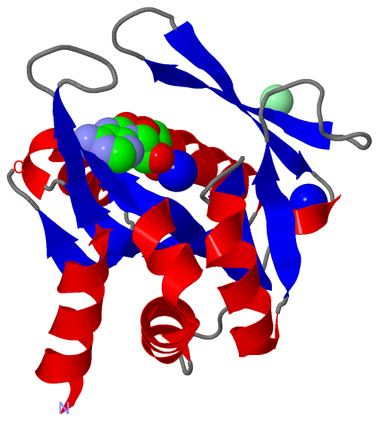
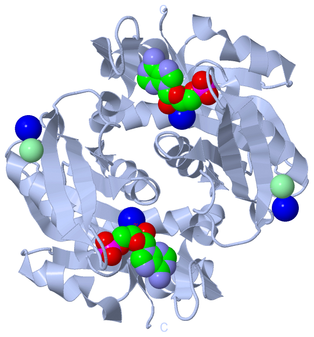
Ligands, Modified Residues, Ions (3, 4)| Asymmetric Unit (3, 4) Biological Unit 1 (1, 2) |
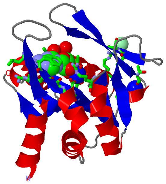
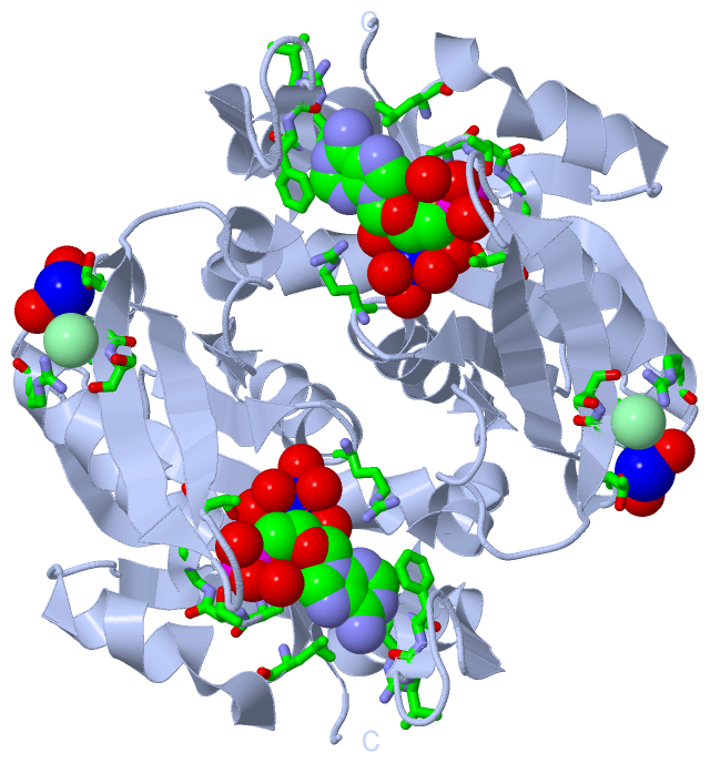
Sites (4, 4)
Asymmetric Unit (4, 4)
|
SS Bonds (0, 0)| (no "SS Bond" information available for 5B6H) |
Cis Peptide Bonds (2, 2)
Asymmetric Unit
|
||||||||||||
SAPs(SNPs)/Variants (0, 0)| (no "SAP(SNP)/Variant" information available for 5B6H) |
PROSITE Motifs (0, 0)| (no "PROSITE Motif" information available for 5B6H) |
Exons (0, 0)| (no "Exon" information available for 5B6H) |
Sequences/Alignments
Asymmetric Unit
Chain A from PDB Type:PROTEIN Length:179
SCOP domains ----------------------------------------------------------------------------------------------------------------------------------------------------------------------------------- SCOP domains
CATH domains ----------------------------------------------------------------------------------------------------------------------------------------------------------------------------------- CATH domains
Pfam domains ----------------------------------------------------------------------------------------------------------------------------------------------------------------------------------- Pfam domains
SAPs(SNPs) ----------------------------------------------------------------------------------------------------------------------------------------------------------------------------------- SAPs(SNPs)
PROSITE ----------------------------------------------------------------------------------------------------------------------------------------------------------------------------------- PROSITE
Transcript ----------------------------------------------------------------------------------------------------------------------------------------------------------------------------------- Transcript
5b6h A 7 KTAQQLKYIKDSIKTIPDYPKAGILFRDVTSLLENPKAYSASIELLSEHYSESGVTKVVGTEARGFLFGAPVALALGVGFVPVRKPGKLPRETISESYELEYGTDTLEIHTDSIQPGDKVLVVDDLLATGGTIEATVKLIRRLGGEVVHAAFIINLPELGGEARLTQQGIHCYSLVSFD 185
16 26 36 46 56 66 76 86 96 106 116 126 136 146 156 166 176
|
||||||||||||||||||||
SCOP Domains (0, 0)| (no "SCOP Domain" information available for 5B6H) |
CATH Domains (0, 0)| (no "CATH Domain" information available for 5B6H) |
Pfam Domains (0, 0)| (no "Pfam Domain" information available for 5B6H) |
Gene Ontology (8, 8)|
Asymmetric Unit(hide GO term definitions) |
Interactive Views
|
|||||||||||||||||||||||||||||||||||||||||||||||||||||||||||||||||||||||||||||||||||||||||||||||||||||||||||||||||||||||||||||||||||||||||||||||||||||||||||||||||||||||||||||||||||
Still Images
|
||||||||||||||||
Databases
|
||||||||||||||||||||||||||||||||||||||||||||||||||||||||||||||||||||||||||||||||||||||||||||||||||||||||||||||||||||||||||||||||||||||||||||||||||||||||||||||||
Analysis Tools
|
|||||||||||||||||||||||||||||||||||||||||||||||||||||||||||||
Entries Sharing at Least One Protein Chain (UniProt ID)
Related Entries Specified in the PDB File
|
|