|
|
|
|
 Description
|
|
Compounds
|
||||||||||||||||||||||||||||||||||||||||||||||||||||||||||||||||||||||||||||||||||||||
Chains, Units
Summary Information (see also Sequences/Alignments below) |
Ligands, Modified Residues, Ions (6, 7)| Asymmetric Unit (6, 7) Biological Unit 1 (2, 4) |
Sites (7, 7)
Asymmetric Unit (7, 7)
|
SS Bonds (0, 0)| (no "SS Bond" information available for 5AUO) |
Cis Peptide Bonds (0, 0)| (no "Cis Peptide Bond" information available for 5AUO) |
SAPs(SNPs)/Variants (0, 0)| (no "SAP(SNP)/Variant" information available for 5AUO) |
PROSITE Motifs (0, 0)| (no "PROSITE Motif" information available for 5AUO) |
Exons (0, 0)| (no "Exon" information available for 5AUO) |
Sequences/Alignments
Asymmetric Unit
Chain A from PDB Type:PROTEIN Length:136
SCOP domains ---------------------------------------------------------------------------------------------------------------------------------------- SCOP domains
CATH domains ---------------------------------------------------------------------------------------------------------------------------------------- CATH domains
Pfam domains ---------------------------------------------------------------------------------------------------------------------------------------- Pfam domains
SAPs(SNPs) ---------------------------------------------------------------------------------------------------------------------------------------- SAPs(SNPs)
PROSITE ---------------------------------------------------------------------------------------------------------------------------------------- PROSITE
Transcript ---------------------------------------------------------------------------------------------------------------------------------------- Transcript
5auo A 1 MHEWALADAIVRTVLDYAQREGASRVKAVRVVLGELQDVAEDIVKFAMEQLFAGTIAEGAEIEFVEEEAVFKCRNCNYEWKLKEVKDKFDERIKEDIHFIPEVVHAFLACPKCGSHDFEVVKGRGVYVAGIKIEKE 136
10 20 30 40 50 60 70 80 90 100 110 120 130
Chain B from PDB Type:PROTEIN Length:237
SCOP domains --------------------------------------------------------------------------------------------------------------------------------------------------------------------------------------------------------------------------------------------- SCOP domains
CATH domains --------------------------------------------------------------------------------------------------------------------------------------------------------------------------------------------------------------------------------------------- CATH domains
Pfam domains --------------------------------------------------------------------------------------------------------------------------------------------------------------------------------------------------------------------------------------------- Pfam domains
SAPs(SNPs) --------------------------------------------------------------------------------------------------------------------------------------------------------------------------------------------------------------------------------------------- SAPs(SNPs)
PROSITE --------------------------------------------------------------------------------------------------------------------------------------------------------------------------------------------------------------------------------------------- PROSITE
Transcript --------------------------------------------------------------------------------------------------------------------------------------------------------------------------------------------------------------------------------------------- Transcript
5auo B 4 IDPREIAINARLEGVKRIIPVVSGKGGVGKSLVSTTLALVLAEKGYRVGLLDLDFHGASDHVILGFEPKEFPEEDRGVVPPTVHGIKFMTIAYYTEDRPTPLRGKEISDALIELLTITRWDELDYLVIDMPPGLGDQLLDVLRFLKRGEFLVVATPSKLSLNVVRKLIELLKEEGHKVIGVVENMKLKDVEKLAEEFGVPYLVGIPFYPDLDAKVGNVEELMKTEFAGKVRELAGRL 248
13 23 33 43 53 63 73 83 93 103 113 123 133 143 153 163 173 183 |201 211 221 231 241
190|
199
|
||||||||||||||||||||
SCOP Domains (0, 0)| (no "SCOP Domain" information available for 5AUO) |
CATH Domains (0, 0)| (no "CATH Domain" information available for 5AUO) |
Pfam Domains (0, 0)| (no "Pfam Domain" information available for 5AUO) |
Gene Ontology (8, 9)|
Asymmetric Unit(hide GO term definitions) |
Interactive Views
|
|||||||||||||||||||||||||||||||||||||||||||||||||||||||||||||||||||||||||||||||||||||||||||||||||||||||||||||||||||||||||||||||||||||||||||||||||||||||||||||||||||||||||||||||||||||||||||||||||||||||||||||||||||||
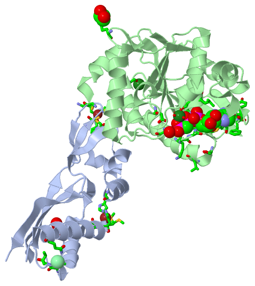
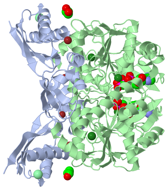
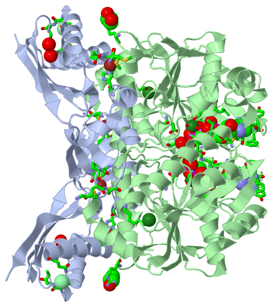
Still Images
|
||||||||||||||||
Databases
|
||||||||||||||||||||||||||||||||||||||||||||||||||||||||||||||||||||||||||||||||||||||||||||||||||||||||||||||||||||||||||||||||||||||||||||||||||||||||||||||||||||||||||||||||||||||||||
Analysis Tools
|
||||||||||||||||||||||||||||||||||||||||||||||||||||||||||||||||||||||||
Entries Sharing at Least One Protein Chain (UniProt ID)
Related Entries Specified in the PDB File
|
|