|
|
|
|
 Description
|
|
Compounds
|
||||||||||||||||||||||||||||||||||||||||||||||||||||
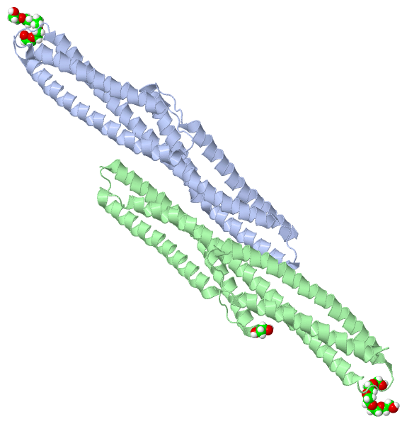
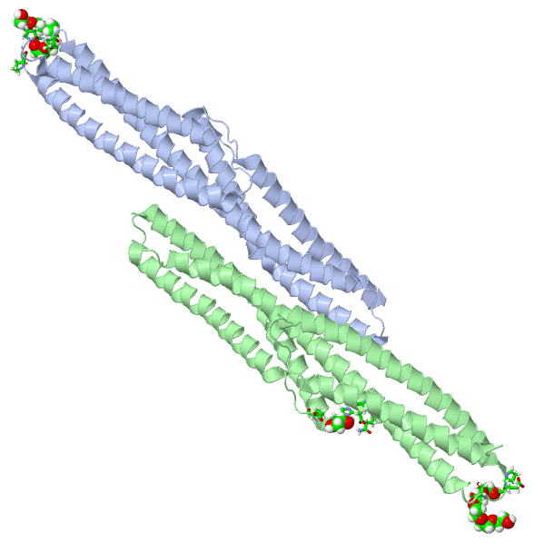
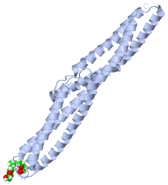
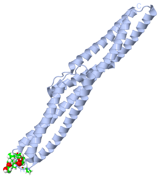
Chains, Units
Summary Information (see also Sequences/Alignments below) |
Ligands, Modified Residues, Ions (2, 3)| Asymmetric Unit (2, 3) Biological Unit 1 (1, 1) Biological Unit 2 (2, 2) |
Sites (3, 3)
Asymmetric Unit (3, 3)
|
SS Bonds (0, 0)| (no "SS Bond" information available for 5J1G) |
Cis Peptide Bonds (0, 0)| (no "Cis Peptide Bond" information available for 5J1G) |
SAPs(SNPs)/Variants (0, 0)| (no "SAP(SNP)/Variant" information available for 5J1G) |
PROSITE Motifs (0, 0)| (no "PROSITE Motif" information available for 5J1G) |
Exons (0, 0)| (no "Exon" information available for 5J1G) |
Sequences/Alignments
Asymmetric Unit
Chain A from PDB Type:PROTEIN Length:228
SCOP domains ------------------------------------------------------------------------------------------------------------------------------------------------------------------------------------------------------------------------------------ SCOP domains
CATH domains ------------------------------------------------------------------------------------------------------------------------------------------------------------------------------------------------------------------------------------ CATH domains
Pfam domains ------------------------------------------------------------------------------------------------------------------------------------------------------------------------------------------------------------------------------------ Pfam domains
SAPs(SNPs) ------------------------------------------------------------------------------------------------------------------------------------------------------------------------------------------------------------------------------------ SAPs(SNPs)
PROSITE ------------------------------------------------------------------------------------------------------------------------------------------------------------------------------------------------------------------------------------ PROSITE
Transcript ------------------------------------------------------------------------------------------------------------------------------------------------------------------------------------------------------------------------------------ Transcript
5j1g A 1004 QEESRCQRCISELKDIRLQLEACETRTVHRLRLPLDKEPARECAQRIAEQQKAQAEVEGLGKGVARLSAEAEKVLALPEPSPAAPTLRSELELTLGKLEQVRSLSAIYLEKLKTISLVIRGTQGAEEVLRAHEEQLKEAQAVPATLPELEATKASLKKLRAQAEAQQPTFDALRDELRGAQEVGERLQQRHGERDVEVERWRERVAQLLERWQAVLAQTDVRQRELEQ 1231
1013 1023 1033 1043 1053 1063 1073 1083 1093 1103 1113 1123 1133 1143 1153 1163 1173 1183 1193 1203 1213 1223
Chain B from PDB Type:PROTEIN Length:229
SCOP domains ------------------------------------------------------------------------------------------------------------------------------------------------------------------------------------------------------------------------------------- SCOP domains
CATH domains ------------------------------------------------------------------------------------------------------------------------------------------------------------------------------------------------------------------------------------- CATH domains
Pfam domains ------------------------------------------------------------------------------------------------------------------------------------------------------------------------------------------------------------------------------------- Pfam domains
SAPs(SNPs) ------------------------------------------------------------------------------------------------------------------------------------------------------------------------------------------------------------------------------------- SAPs(SNPs)
PROSITE ------------------------------------------------------------------------------------------------------------------------------------------------------------------------------------------------------------------------------------- PROSITE
Transcript ------------------------------------------------------------------------------------------------------------------------------------------------------------------------------------------------------------------------------------- Transcript
5j1g B 1005 EESRCQRCISELKDIRLQLEACETRTVHRLRLPLDKEPARECAQRIAEQQKAQAEVEGLGKGVARLSAEAEKVLALPEPSPAAPTLRSELELTLGKLEQVRSLSAIYLEKLKTISLVIRGTQGAEEVLRAHEEQLKEAQAVPATLPELEATKASLKKLRAQAEAQQPTFDALRDELRGAQEVGERLQQRHGERDVEVERWRERVAQLLERWQAVLAQTDVRQRELEQLG 1233
1014 1024 1034 1044 1054 1064 1074 1084 1094 1104 1114 1124 1134 1144 1154 1164 1174 1184 1194 1204 1214 1224
|
||||||||||||||||||||
SCOP Domains (0, 0)| (no "SCOP Domain" information available for 5J1G) |
CATH Domains (0, 0)| (no "CATH Domain" information available for 5J1G) |
Pfam Domains (0, 0)| (no "Pfam Domain" information available for 5J1G) |
Gene Ontology (20, 20)|
Asymmetric Unit(hide GO term definitions) |
Interactive Views
|
||||||||||||||||||||||||||||||||||||||||||||||||||||||||||||||||||||||||||||||||||||||||||||||||||||||||||||||||||||||||||||||||||||||||||||||||||||||||||||||||||
Still Images
|
||||||||||||||||
Databases
|
||||||||||||||||||||||||||||||||||||||||||||||||||||||||||||||||||||||||||||||||||||||||||||||||||||||||||||||||||||||||||||||||||||||||||||||||||||||||||||||||
Analysis Tools
|
|||||||||||||||||||||||||||||||||||||||||||||||||||||||||||||
Entries Sharing at Least One Protein Chain (UniProt ID)
Related Entries Specified in the PDB File
|
|