Show PDB file:   
         Plain Text   HTML   (compressed file size)
QuickSearch:   
by PDB,NDB,UniProt,PROSITE Code or Search Term(s)  
(-)Asymmetric Unit
(-)Asym. Unit - sites
(-)Biological Unit 1
(-)Biol. Unit 1 - sites
collapse expand < >
Image Asymmetric Unit
Asymmetric Unit  (Jmol Viewer)
Image Asym. Unit - sites
Asym. Unit - sites  (Jmol Viewer)
Image Biological Unit 1
Biological Unit 1  (Jmol Viewer)
Image Biol. Unit 1 - sites
Biol. Unit 1 - sites  (Jmol Viewer)

(-) Description

Title :  CRYSTAL STRUCTURE OF M. MUSCULUS PROTEIN ARGININE METHYLTRANSFERASE PRMT6 WITH SAH AND MAGNESIUM
 
Authors :  L. Bonnefond, J. Cavarelli
Date :  14 Dec 15  (Deposition) - 18 Jan 17  (Release) - 18 Jan 17  (Revision)
Method :  X-RAY DIFFRACTION
Resolution :  1.90
Chains :  Asym. Unit :  A
Biol. Unit 1:  A  (2x)
Keywords :  Transferase, S-Adenosyl-L-Methionine (Keyword Search: [Gene Ontology, PubMed, Web (Google))
 
Reference :  L. Bonnefond, V. Cura, N. Troffer-Charlier, J. Cavarelli
Crystal Strcutures Of Prmt6 In Complex With Sah In Alternative Conformations
To Be Published
PubMed: search

(-) Compounds

Molecule 1 - PROTEIN ARGININE N-METHYLTRANSFERASE 6
    ChainsA
    EC Number2.1.1.-, 2.1.1.125
    EngineeredYES
    Expression SystemESCHERICHIA COLI
    Expression System PlasmidPNEA-VH
    Expression System StrainBL21(DE3)
    Expression System Taxid469008
    Expression System VectorPET15 DERIVATIVE
    Expression System Vector TypePLASMID
    Organism CommonHOUSE MOUSE
    Organism ScientificMUS MUSCULUS
    Organism Taxid10090
    SynonymHISTONE-ARGININE N-METHYLTRANSFERASE PRMT6

 Structural Features

(-) Chains, Units

  1
Asymmetric Unit A
Biological Unit 1 (2x)A

Summary Information (see also Sequences/Alignments below)
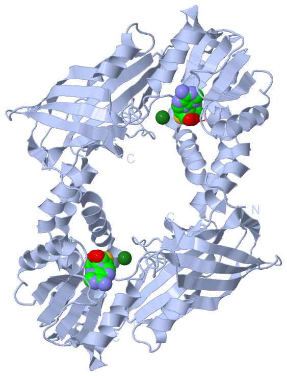
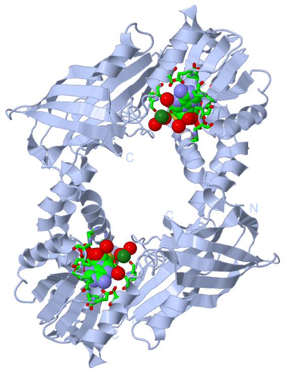
(-) Ligands, Modified Residues, Ions  (2, 2)

Asymmetric Unit (2, 2)
No.NameCountTypeFull Name
1MG1Ligand/IonMAGNESIUM ION
2SAH1Ligand/IonS-ADENOSYL-L-HOMOCYSTEINE
Biological Unit 1 (1, 2)
No.NameCountTypeFull Name
1MG-1Ligand/IonMAGNESIUM ION
2SAH2Ligand/IonS-ADENOSYL-L-HOMOCYSTEINE

(-) Sites  (2, 2)

Asymmetric Unit (2, 2)
No.NameEvidenceResiduesDescription
1AC1SOFTWAREHIS A:60 , MET A:63 , ARG A:69 , GLY A:93 , ALA A:94 , LEU A:99 , GLU A:115 , ALA A:116 , SER A:117 , GLY A:141 , PRO A:142 , VAL A:143 , GLU A:144 , GLU A:158 , MET A:169 , SER A:172 , HOH A:2008 , HOH A:2036 , HOH A:2037 , HOH A:2109 , HOH A:2247BINDING SITE FOR RESIDUE SAH A1378
2AC2SOFTWAREGLU A:158 , MET A:160 , HOH A:2014 , HOH A:2015 , HOH A:2092 , HOH A:2096 , HOH A:2097BINDING SITE FOR RESIDUE MG A1379

(-) SS Bonds  (1, 1)

Asymmetric Unit
No.Residues
1A:53 -A:232

(-) Cis Peptide Bonds  (1, 1)

Asymmetric Unit
No.Residues
1Leu A:188 -Pro A:189

 Sequence-Structure Mapping

(-) SAPs(SNPs)/Variants  (0, 0)

(no "SAP(SNP)/Variant" information available for 5FQO)

(-) PROSITE Motifs  (0, 0)

(no "PROSITE Motif" information available for 5FQO)

(-) Exons   (0, 0)

(no "Exon" information available for 5FQO)

(-) Sequences/Alignments

Asymmetric Unit
   Reformat: Number of residues per line =  ('0' or empty: single-line sequence representation)
  Number of residues per labelling interval =   
  UniProt sequence: complete  aligned part    
   Show mapping: SCOP domains CATH domains Pfam domains Secondary structure (by author)
SAPs(SNPs) PROSITE motifs Exons
(details for a mapped element are shown in a popup box when the mouse pointer rests over it)
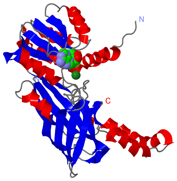
Chain A from PDB  Type:PROTEIN  Length:324
                                                                                                                                                                                                                                                                                                                                                                    
               SCOP domains ------------------------------------------------------------------------------------------------------------------------------------------------------------------------------------------------------------------------------------------------------------------------------------------------------------------------------------ SCOP domains
               CATH domains ------------------------------------------------------------------------------------------------------------------------------------------------------------------------------------------------------------------------------------------------------------------------------------------------------------------------------------ CATH domains
               Pfam domains ------------------------------------------------------------------------------------------------------------------------------------------------------------------------------------------------------------------------------------------------------------------------------------------------------------------------------------ Pfam domains
         Sec.struct. author ....hhhhhhhhhhhhhhhhhhhhhhhhhhhhhhhhh..eeeee....hhhhhhhhhh...eeeeee...hhhhhhhhhhhh.....eeeee...........eeeee............hhhhhhhhhhhheeeeeeee..eeeeeeeee.hhhhhhhhhhhhhhhhhhh....hhhhhhhhhhhh..eeeee..hhhh.....eeeeeee....hhhhhhhhheeeeeeee....eeeeeeeeeeeeee...eeee...........eeeeeeeeeeee....eeeeeeeeee.......eeeeeeeee.....eeeeeee. Sec.struct. author
                 SAPs(SNPs) ------------------------------------------------------------------------------------------------------------------------------------------------------------------------------------------------------------------------------------------------------------------------------------------------------------------------------------ SAPs(SNPs)
                    PROSITE ------------------------------------------------------------------------------------------------------------------------------------------------------------------------------------------------------------------------------------------------------------------------------------------------------------------------------------ PROSITE
                 Transcript ------------------------------------------------------------------------------------------------------------------------------------------------------------------------------------------------------------------------------------------------------------------------------------------------------------------------------------ Transcript
                 5fqo A  49 LYYECYSDVSVHEEMIADQVRTEAYRLGILKNWAALRGKTVLDVGAGTGILSIFCAQAGARRVYAVEASAIWQQAREVVRLNGLEDRVHVLPGPVETVELPERVDAIVSEWMGYGLLHESMLSSVLHARTKWLKEGGLLLPASAELFVAPISDQMLEWRLGFWSQVKQHYGVDMSCMESFATRCLMGHSEIVVQDLSGEDVLARPQRFAQLELARAGLEQELEAGVGGRFRCSCYGSAPLHGFAVWFQVTFPGPLVLSTSPLHPATHWKQALLYLNEPVPVEQDTDISGEITLLPSPDNPRRLRILLRYKVGDHEEKTKDFAME 377
                                    58        68        78        88        98       108       118       128       138       148       158       168       178       188       198       208       218       228       238       248       258       268       278       288       298  ||   313       323       333       343       353       363       373    
                                                                                                                                                                                                                                                                                      301|                                                                      
                                                                                                                                                                                                                                                                                       307                                                                      

   Legend:   → Mismatch (orange background)
  - → Gap (green background, '-', border residues have a numbering label)
    → Modified Residue (blue background, lower-case, 'x' indicates undefined single-letter code, labelled with number + name)
  x → Chemical Group (purple background, 'x', labelled with number + name, e.g. ACE or NH2)
  extra numbering lines below/above indicate numbering irregularities and modified residue names etc., number ends below/above '|'

 Classification and Annotation

(-) SCOP Domains  (0, 0)

(no "SCOP Domain" information available for 5FQO)

(-) CATH Domains  (0, 0)

(no "CATH Domain" information available for 5FQO)

(-) Pfam Domains  (0, 0)

(no "Pfam Domain" information available for 5FQO)

(-) Gene Ontology  (28, 28)

Asymmetric Unit(hide GO term definitions)

 Visualization

(-) Interactive Views

Asymmetric Unit
  Complete Structure
    Jena3D(integrated viewing of ligand, site, SAP, PROSITE, SCOP information)
    WebMol | AstexViewer[tm]@PDBe
(Java Applets, require no local installation except for Java; loading may be slow)
    STRAP
(Java WebStart application, automatic local installation, requires Java; full application with system access!)
    RasMol
(require local installation)
    Molscript (VRML)
(requires installation of a VRML viewer; select preferred view via VRML and generate a mono or stereo PDF format file)
 
  Ligands, Modified Residues, Ions
    MG  [ RasMol | Jena3D ]  +environment [ RasMol | Jena3D ]
    SAH  [ RasMol | Jena3D ]  +environment [ RasMol | Jena3D ]
 
  Sites
    AC1  [ RasMol ]  +environment [ RasMol ]
    AC2  [ RasMol ]  +environment [ RasMol ]
 
  Cis Peptide Bonds
    Leu A:188 - Pro A:189   [ RasMol ]  
 
Biological Unit
  Complete Structure
    Biological Unit 1  [ Jena3D ]

(-) Still Images

Jmol
  protein: cartoon or spacefill or dots and stick; nucleic acid: cartoon and stick; ligands: spacefill; active site: stick
Molscript
  protein, nucleic acid: cartoon; ligands: spacefill; active site: ball and stick

 Databases and Analysis Tools

(-) Databases

Access by PDB/NDB ID
  5fqo
    Family and Domain InformationProDom | SYSTERS
    General Structural InformationGlycoscienceDB | MMDB | NDB | OCA | PDB | PDBe | PDBj | PDBsum | PDBWiki | PQS | PROTEOPEDIA
    Orientation in MembranesOPM
    Protein SurfaceSURFACE
    Secondary StructureDSSP (structure derived) | HSSP (homology derived)
    Structural GenomicsGeneCensus
    Structural NeighboursCE | VAST
    Structure ClassificationCATH | Dali | SCOP
    Validation and Original DataBMRB Data View | BMRB Restraints Grid | EDS | PROCHECK | RECOORD | WHAT_CHECK
 
Access by UniProt ID/Accession number
  ANM6_MOUSE | Q6NZB1
    Comparative Protein Structure ModelsModBase
    Genomic InformationEnsembl
    Protein-protein InteractionDIP
    Sequence, Family and Domain InformationInterPro | Pfam | SMART | UniProtKB/SwissProt
 
Access by Enzyme Classificator   (EC Number)
  2.1.1.-
    General Enzyme InformationBRENDA | EC-PDB | Enzyme | IntEnz
    PathwayKEGG | MetaCyc
  2.1.1.125
    General Enzyme InformationBRENDA | EC-PDB | Enzyme | IntEnz
    PathwayKEGG | MetaCyc
 
Access by Disease Identifier   (MIM ID)
  (no 'MIM ID' available)
    Disease InformationOMIM
 
Access by GenAge ID
  (no 'GenAge ID' available)
    Age Related InformationGenAge

(-) Analysis Tools

Access by PDB/NDB ID
    Domain InformationXDom
    Interatomic Contacts of Structural UnitsCSU
    Ligand-protein ContactsLPC
    Protein CavitiescastP
    Sequence and Secondary StructurePDBCartoon
    Structure AlignmentSTRAP(Java WebStart application, automatic local installation, requires Java; full application with system access!)
    Structure and Sequence BrowserSTING
 
Access by UniProt ID/Accession number
  ANM6_MOUSE | Q6NZB1
    Protein Disorder PredictionDisEMBL | FoldIndex | GLOBPLOT (for more information see DisProt)

 Related Entries

(-) Entries Sharing at Least One Protein Chain (UniProt ID)

UniProtKB/Swiss-Prot
        ANM6_MOUSE | Q6NZB14c03 4c04 4c05 4c06 4c07 4c08 5fqn

(-) Related Entries Specified in the PDB File

5fqn CRYSTAL STRUCTURE OF M. MUSCULUS PROTEIN ARGININE METHYLTRANSFERASE PRMT6 WITH SAH AT 1.65 ANGSTROMS