Show PDB file:   
         Plain Text   HTML   (compressed file size)
QuickSearch:   
by PDB,NDB,UniProt,PROSITE Code or Search Term(s)  
(-)Asymmetric Unit
(-)Asym. Unit - sites
(-)Biological Unit 1
(-)Biol. Unit 1 - sites
(-)Biological Unit 2
collapse expand < >
Image Asymmetric Unit
Asymmetric Unit  (Jmol Viewer)
Image Asym. Unit - sites
Asym. Unit - sites  (Jmol Viewer)
Image Biological Unit 1
Biological Unit 1  (Jmol Viewer)
Image Biol. Unit 1 - sites
Biol. Unit 1 - sites  (Jmol Viewer)
Image Biological Unit 2
Biological Unit 2  (Jmol Viewer)

(-) Description

Title :  CRYSTAL STRUCTURE OF C-TERMINAL DOMAIN OF THE HUMAN DNA PRIMASE LARGE SUBUNIT WITH BOUND DNA TEMPLATE/RNA PRIMER AND MANGANESE ION
 
Authors :  T. H. Tahirov, A. G. Baranovskiy, N. D. Babayeva
Date :  28 Nov 15  (Deposition) - 23 Mar 16  (Release) - 01 Jun 16  (Revision)
Method :  X-RAY DIFFRACTION
Resolution :  3.00
Chains :  Asym. Unit :  A,B,C,D,E,F
Biol. Unit 1:  A,C,D  (1x)
Biol. Unit 2:  B,E,F  (1x)
Keywords :  Tranferase-Dna-Rna Complex, Dna Primase, Large Subunit, Iron-Sulfur Cluster, Rna, Dna, Primer, Template, Triphosphate, Initiation Site, Manganese, Replication-Dna-Rna Complex (Keyword Search: [Gene Ontology, PubMed, Web (Google))
 
Reference :  A. G. Baranovskiy, N. D. Babayeva, Y. Zhang, J. Gu, Y. Suwa, Y. I. Pavlov T. H. Tahirov
Mechanism Of Concerted Rna-Dna Primer Synthesis By The Huma Primosome.
J. Biol. Chem. V. 291 10006 2016
PubMed-ID: 26975377  |  Reference-DOI: 10.1074/JBC.M116.717405

(-) Compounds

Molecule 1 - DNA PRIMASE LARGE SUBUNIT
    ChainsA, B
    EC Number2.7.7.-
    EngineeredYES
    Expression SystemESCHERICHIA COLI
    Expression System Taxid562
    FragmentUNP RESIDUES 266-456
    GenePRIM2, PRIM2A
    Organism CommonHUMAN
    Organism ScientificHOMO SAPIENS
    Organism Taxid9606
    SynonymDNA PRIMASE 58 KDA SUBUNIT,P58
 
Molecule 2 - RNA (5'-R(P*GP*GP*CP*GP*GP*C)-3')
    ChainsC, E
    EngineeredYES
    Organism ScientificHOMO SAPIENS
    Organism Taxid9606
    Other DetailsCONTAINS 5'-TRIPHOSPHATE
    SyntheticYES
 
Molecule 3 - DNA (5'-D(*GP*CP*CP*GP*CP*CP*AP*AP*CP*AP*TP*A)-3')
    ChainsD, F
    EngineeredYES
    Organism ScientificHOMO SAPIENS
    Organism Taxid9606
    SyntheticYES

 Structural Features

(-) Chains, Units

  123456
Asymmetric Unit ABCDEF
Biological Unit 1 (1x)A CD  
Biological Unit 2 (1x) B  EF

Summary Information (see also Sequences/Alignments below)

(-) Ligands, Modified Residues, Ions  (3, 6)

Asymmetric Unit (3, 6)
No.NameCountTypeFull Name
1GTP2Mod. NucleotideGUANOSINE-5'-TRIPHOSPHATE
2MN2Ligand/IonMANGANESE (II) ION
3SF42Ligand/IonIRON/SULFUR CLUSTER
Biological Unit 1 (2, 2)
No.NameCountTypeFull Name
1GTP1Mod. NucleotideGUANOSINE-5'-TRIPHOSPHATE
2MN-1Ligand/IonMANGANESE (II) ION
3SF41Ligand/IonIRON/SULFUR CLUSTER
Biological Unit 2 (2, 2)
No.NameCountTypeFull Name
1GTP1Mod. NucleotideGUANOSINE-5'-TRIPHOSPHATE
2MN-1Ligand/IonMANGANESE (II) ION
3SF41Ligand/IonIRON/SULFUR CLUSTER

(-) Sites  (5, 5)

Asymmetric Unit (5, 5)
No.NameEvidenceResiduesDescription
1AC1SOFTWAREPRO A:286 , CYS A:287 , CYS A:367 , CYS A:384 , CYS A:424 , LEU A:441binding site for residue SF4 A 501
2AC2SOFTWAREPRO B:285 , PRO B:286 , CYS B:287 , CYS B:367 , CYS B:384 , PHE B:386 , CYS B:424 , PRO B:444binding site for residue SF4 B 501
3AC3SOFTWAREGTP C:1binding site for residue MN C 101
4AC4SOFTWARETYR B:345 , GTP E:1binding site for residue MN E 101
5AC5SOFTWAREHIS B:300 , ARG B:302 , HIS B:303 , ARG B:306 , TYR B:345 , C E:3 , MN E:101 , DC F:5 , DC F:6 , DA F:7binding site for Di-nucleotide GTP E 1 and G E 2

(-) SS Bonds  (0, 0)

(no "SS Bond" information available for 5F0S)

(-) Cis Peptide Bonds  (0, 0)

(no "Cis Peptide Bond" information available for 5F0S)

 Sequence-Structure Mapping

(-) SAPs(SNPs)/Variants  (0, 0)

(no "SAP(SNP)/Variant" information available for 5F0S)

(-) PROSITE Motifs  (0, 0)

(no "PROSITE Motif" information available for 5F0S)

(-) Exons   (0, 0)

(no "Exon" information available for 5F0S)

(-) Sequences/Alignments

Asymmetric Unit
   Reformat: Number of residues per line =  ('0' or empty: single-line sequence representation)
  Number of residues per labelling interval =   
  UniProt sequence: complete  aligned part    
   Show mapping: SCOP domains CATH domains Pfam domains Secondary structure (by author)
SAPs(SNPs) PROSITE motifs Exons
(details for a mapped element are shown in a popup box when the mouse pointer rests over it)
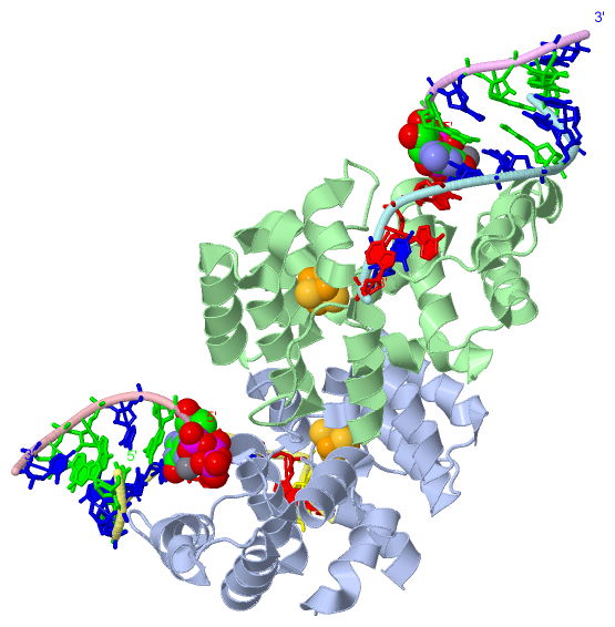
Chain A from PDB  Type:PROTEIN  Length:187
                                                                                                                                                                                                                           
               SCOP domains ------------------------------------------------------------------------------------------------------------------------------------------------------------------------------------------- SCOP domains
               CATH domains ------------------------------------------------------------------------------------------------------------------------------------------------------------------------------------------- CATH domains
               Pfam domains ------------------------------------------------------------------------------------------------------------------------------------------------------------------------------------------- Pfam domains
         Sec.struct. author ..hhhhhhhhhhhh.hhhhhhhhhhhhhhh..hhhhhhhhhhhhhhhh.hhhhhhhhhhhhhhh...hhhhhhhhhhhhhhhhh............hhhhhhhh..........hhhhhhhhhhhhhhhhhh..hhhhhhhhhhhhhh.hhhhhhhhhhhhhh..........hhhhhhhhhhhhhh Sec.struct. author
                 SAPs(SNPs) ------------------------------------------------------------------------------------------------------------------------------------------------------------------------------------------- SAPs(SNPs)
                    PROSITE ------------------------------------------------------------------------------------------------------------------------------------------------------------------------------------------- PROSITE
                 Transcript ------------------------------------------------------------------------------------------------------------------------------------------------------------------------------------------- Transcript
                 5f0s A 270 KISLDQIDLLSTKSFPPCMRQLHKALRENHHLRHGGRMQYGLFLKGIGLTLEQALQFWKQEFIKGKMDPDKFDKGYSYNIRHSFGKEGKRTDYTPFSCLKIILSNPPSQGDYHGCPFRHSDPELLKQKLQSYKISPGGISQILDLVKGTHYQVACQKYFEMIHNVDDCGFSLNHPNQFFCESQRILN 456
                                   279       289       299       309       319       329       339       349       359       369       379       389       399       409       419       429       439       449       

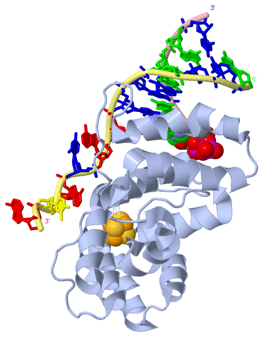
Chain B from PDB  Type:PROTEIN  Length:185
                                                                                                                                                                                                                         
               SCOP domains ----------------------------------------------------------------------------------------------------------------------------------------------------------------------------------------- SCOP domains
               CATH domains ----------------------------------------------------------------------------------------------------------------------------------------------------------------------------------------- CATH domains
               Pfam domains ----------------------------------------------------------------------------------------------------------------------------------------------------------------------------------------- Pfam domains
         Sec.struct. author .hhhhhhhhhhhh.hhhhhhhhhhhhhhh..hhhhhhhhhhhhhhh..hhhhhhhhhhhhhhh...hhhhhhhhhhhhhhhhh............hhhhhhhh..........hhhhhhhhhhhhhhhhhh..hhhhhhhhhhhhhh.hhhhhhhhhhhhhh..........hhhhhhhhhhhhh Sec.struct. author
                 SAPs(SNPs) ----------------------------------------------------------------------------------------------------------------------------------------------------------------------------------------- SAPs(SNPs)
                    PROSITE ----------------------------------------------------------------------------------------------------------------------------------------------------------------------------------------- PROSITE
                 Transcript ----------------------------------------------------------------------------------------------------------------------------------------------------------------------------------------- Transcript
                 5f0s B 271 ISLDQIDLLSTKSFPPCMRQLHKALRENHHLRHGGRMQYGLFLKGIGLTLEQALQFWKQEFIKGKMDPDKFDKGYSYNIRHSFGKEGKRTDYTPFSCLKIILSNPPSQGDYHGCPFRHSDPELLKQKLQSYKISPGGISQILDLVKGTHYQVACQKYFEMIHNVDDCGFSLNHPNQFFCESQRIL 455
                                   280       290       300       310       320       330       340       350       360       370       380       390       400       410       420       430       440       450     

Chain C from PDB  Type:RNA  Length:6
                                      
                 5f0s C   1 xGCGGC   6
                            |     
                            1-GTP 

Chain D from PDB  Type:DNA  Length:12
                                            
                 5f0s D   1 GCCGCCAACATA  12
                                    10  

Chain E from PDB  Type:RNA  Length:6
                                      
                 5f0s E   1 xGCGGC   6
                            |     
                            1-GTP 

Chain F from PDB  Type:DNA  Length:10
                                          
                 5f0s F   1 GCCGCCAACA  10
                                    10

   Legend:   → Mismatch (orange background)
  - → Gap (green background, '-', border residues have a numbering label)
    → Modified Residue (blue background, lower-case, 'x' indicates undefined single-letter code, labelled with number + name)
  x → Chemical Group (purple background, 'x', labelled with number + name, e.g. ACE or NH2)
  extra numbering lines below/above indicate numbering irregularities and modified residue names etc., number ends below/above '|'

 Classification and Annotation

(-) SCOP Domains  (0, 0)

(no "SCOP Domain" information available for 5F0S)

(-) CATH Domains  (0, 0)

(no "CATH Domain" information available for 5F0S)

(-) Pfam Domains  (0, 0)

(no "Pfam Domain" information available for 5F0S)

(-) Gene Ontology  (16, 16)

Asymmetric Unit(hide GO term definitions)

 Visualization

(-) Interactive Views

Asymmetric Unit
  Complete Structure
    Jena3D(integrated viewing of ligand, site, SAP, PROSITE, SCOP information)
    WebMol | AstexViewer[tm]@PDBe
(Java Applets, require no local installation except for Java; loading may be slow)
    STRAP
(Java WebStart application, automatic local installation, requires Java; full application with system access!)
    RasMol
(require local installation)
    Molscript (VRML)
(requires installation of a VRML viewer; select preferred view via VRML and generate a mono or stereo PDF format file)
 
  Ligands, Modified Residues, Ions
    GTP  [ RasMol | Jena3D ]  +environment [ RasMol | Jena3D ]
    MN  [ RasMol | Jena3D ]  +environment [ RasMol | Jena3D ]
    SF4  [ RasMol | Jena3D ]  +environment [ RasMol | Jena3D ]
 
  Sites
    AC1  [ RasMol ]  +environment [ RasMol ]
    AC2  [ RasMol ]  +environment [ RasMol ]
    AC3  [ RasMol ]  +environment [ RasMol ]
    AC4  [ RasMol ]  +environment [ RasMol ]
    AC5  [ RasMol ]  +environment [ RasMol ]
 
  Cis Peptide Bonds
(no "Cis Peptide Bonds" information available for 5f0s)
 
Biological Units
  Complete Structure
    Biological Unit 1  [ Jena3D ]
    Biological Unit 2  [ Jena3D ]

(-) Still Images

Jmol
  protein: cartoon or spacefill or dots and stick; nucleic acid: cartoon and stick; ligands: spacefill; active site: stick
Molscript
  protein, nucleic acid: cartoon; ligands: spacefill; active site: ball and stick

 Databases and Analysis Tools

(-) Databases

Access by PDB/NDB ID
  5f0s
    Family and Domain InformationProDom | SYSTERS
    General Structural InformationGlycoscienceDB | MMDB | NDB | OCA | PDB | PDBe | PDBj | PDBsum | PDBWiki | PQS | PROTEOPEDIA
    Orientation in MembranesOPM
    Protein SurfaceSURFACE
    Secondary StructureDSSP (structure derived) | HSSP (homology derived)
    Structural GenomicsGeneCensus
    Structural NeighboursCE | VAST
    Structure ClassificationCATH | Dali | SCOP
    Validation and Original DataBMRB Data View | BMRB Restraints Grid | EDS | PROCHECK | RECOORD | WHAT_CHECK
 
Access by UniProt ID/Accession number
  PRI2_HUMAN | P49643
    Comparative Protein Structure ModelsModBase
    Genomic InformationEnsembl
    Protein-protein InteractionDIP
    Sequence, Family and Domain InformationInterPro | Pfam | SMART | UniProtKB/SwissProt
 
Access by Enzyme Classificator   (EC Number)
  2.7.7.-
    General Enzyme InformationBRENDA | EC-PDB | Enzyme | IntEnz
    PathwayKEGG | MetaCyc
 
Access by Disease Identifier   (MIM ID)
  (no 'MIM ID' available)
    Disease InformationOMIM
 
Access by GenAge ID
  (no 'GenAge ID' available)
    Age Related InformationGenAge

(-) Analysis Tools

Access by PDB/NDB ID
    Domain InformationXDom
    Interatomic Contacts of Structural UnitsCSU
    Ligand-protein ContactsLPC
    Protein CavitiescastP
    Sequence and Secondary StructurePDBCartoon
    Structure AlignmentSTRAP(Java WebStart application, automatic local installation, requires Java; full application with system access!)
    Structure and Sequence BrowserSTING
 
Access by UniProt ID/Accession number
  PRI2_HUMAN | P49643
    Protein Disorder PredictionDisEMBL | FoldIndex | GLOBPLOT (for more information see DisProt)

 Related Entries

(-) Entries Sharing at Least One Protein Chain (UniProt ID)

UniProtKB/Swiss-Prot
        PRI2_HUMAN | P496433l9q 3q36 4bpu 4bpw 4bpx 4rr2 5dqo 5exr 5f0q 5i7m

(-) Related Entries Specified in the PDB File

5f0q CONTAINS MG INSTEAD OF MN