Show PDB file:   
         Plain Text   HTML   (compressed file size)
QuickSearch:   
by PDB,NDB,UniProt,PROSITE Code or Search Term(s)  
(-)Asymmetric Unit
(-)Asym. Unit - sites
(-)Biological Unit 1
(-)Biol. Unit 1 - sites
(-)Biological Unit 2
collapse expand < >
Image Asymmetric Unit
Asymmetric Unit  (Jmol Viewer)
Image Asym. Unit - sites
Asym. Unit - sites  (Jmol Viewer)
Image Biological Unit 1
Biological Unit 1  (Jmol Viewer)
Image Biol. Unit 1 - sites
Biol. Unit 1 - sites  (Jmol Viewer)
Image Biological Unit 2
Biological Unit 2  (Jmol Viewer)

(-) Description

Title :  CRYSTAL STRUCTURE OF THE LECTIN DOMAIN OF PAPG FROM E. COLI BI47 IN COMPLEX WITH 4-METHOXYPHENYL BETA-D-GALABIOSE IN SPACE GROUP P212121
 
Authors :  R. P. Jakob, G. Navarra, P. Zihlmann, K. Stangier, R. C. Preston, S. Rabb T. Maier, B. Ernst
Date :  31 Mar 15  (Deposition) - 13 Apr 16  (Release) - 13 Apr 16  (Revision)
Method :  X-RAY DIFFRACTION
Resolution :  1.45
Chains :  Asym. Unit :  A,B
Biol. Unit 1:  A  (1x)
Biol. Unit 2:  B  (1x)
Keywords :  Upec, Urinary Tract Infection, Fimbrial Adhesin, Adhesin, Type I Pili, Papg, Carbohydrate Binding, Sugar Binding Protein (Keyword Search: [Gene Ontology, PubMed, Web (Google))
 
Reference :  G. Navarra, P. Zihlmann, R. P. Jakob, K. Stangier, R. C. Preston, S. Rabbani, T. Maier, B. Ernst
An Unexpected Way Nature Improves Solvation Entropy
To Be Published
PubMed: search

(-) Compounds

Molecule 1 - PAPG, LECTIN DOMAIN
    ChainsA, B
    EngineeredYES
    Expression SystemESCHERICHIA COLI BL21(DE3)
    Expression System PlasmidPET-22B
    Expression System Taxid469008
    Expression System VariantADA494
    FragmentRESIDUES 20-216
    GeneG801_04654, G801_04690
    Organism ScientificESCHERICHIA COLI
    Organism Taxid562
    Strain4-5674999

 Structural Features

(-) Chains, Units

  12
Asymmetric Unit AB
Biological Unit 1 (1x)A 
Biological Unit 2 (1x) B

Summary Information (see also Sequences/Alignments below)

(-) Ligands, Modified Residues, Ions  (4, 8)

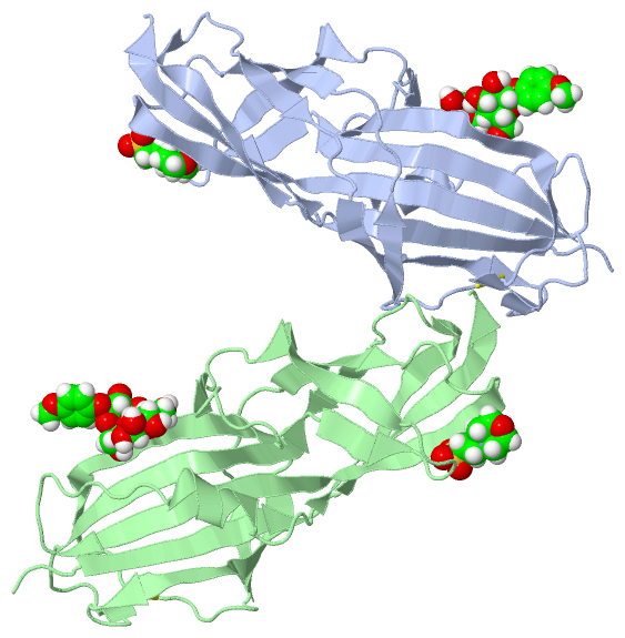
Asymmetric Unit (4, 8)
No.NameCountTypeFull Name
14KS2Ligand/Ion4-METHOXYPHENOL
2GAL2Ligand/IonBETA-D-GALACTOSE
3GLA2Ligand/IonALPHA D-GALACTOSE
4MES2Ligand/Ion2-(N-MORPHOLINO)-ETHANESULFONIC ACID
Biological Unit 1 (4, 4)
No.NameCountTypeFull Name
14KS1Ligand/Ion4-METHOXYPHENOL
2GAL1Ligand/IonBETA-D-GALACTOSE
3GLA1Ligand/IonALPHA D-GALACTOSE
4MES1Ligand/Ion2-(N-MORPHOLINO)-ETHANESULFONIC ACID
Biological Unit 2 (4, 4)
No.NameCountTypeFull Name
14KS1Ligand/Ion4-METHOXYPHENOL
2GAL1Ligand/IonBETA-D-GALACTOSE
3GLA1Ligand/IonALPHA D-GALACTOSE
4MES1Ligand/Ion2-(N-MORPHOLINO)-ETHANESULFONIC ACID

(-) Sites  (4, 4)

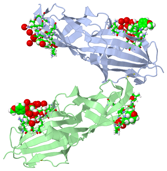
Asymmetric Unit (4, 4)
No.NameEvidenceResiduesDescription
1AC1SOFTWARELYS A:73 , ASN A:74 , LEU A:146 , GLY A:147 , ASP A:148 , TYR A:149 , SER A:150 , HOH A:317 , HOH A:462 , HOH A:485 , HOH A:526binding site for residue MES A 201
2AC2SOFTWARELYS B:73 , ASN B:74 , LEU B:146 , GLY B:147 , ASP B:148 , TYR B:149 , SER B:150binding site for residue MES B 201
3AC3SOFTWAREGLU A:59 , GLU A:91 , LYS A:103 , GLY A:104 , TRP A:107 , ARG A:170 , LYS A:172 , HOH A:305 , HOH A:325 , HOH A:333 , HOH A:342 , HOH A:425 , HOH A:493 , GLN B:16 , ASP B:148binding site for Poly-Saccharide residues 4KS A 202 through GLA A 204
4AC4SOFTWAREGLN A:16 , GLY A:18 , ASN A:19 , GLU A:59 , GLU A:91 , LYS A:103 , GLY A:104 , TRP A:107 , ARG A:170 , LYS A:172 , LEU A:190 , LYS A:192 , HOH A:305 , HOH A:325 , HOH A:333 , HOH A:335 , HOH A:342 , HOH A:408 , HOH A:425 , HOH A:493 , GLN B:16 , GLU B:59 , GLU B:91 , LYS B:103 , GLY B:104 , TRP B:107 , ASP B:148 , ARG B:170 , LYS B:172 , HOH B:333 , HOH B:346 , HOH B:350 , HOH B:382 , HOH B:387 , HOH B:442 , HOH B:452binding site for Poly-Saccharide residues 4KS B 202 through GLA B 204

(-) SS Bonds  (2, 2)

Asymmetric Unit
No.Residues
1A:44 -A:118
2B:44 -B:118

(-) Cis Peptide Bonds  (0, 0)

(no "Cis Peptide Bond" information available for 4Z3G)

 Sequence-Structure Mapping

(-) SAPs(SNPs)/Variants  (0, 0)

(no "SAP(SNP)/Variant" information available for 4Z3G)

(-) PROSITE Motifs  (0, 0)

(no "PROSITE Motif" information available for 4Z3G)

(-) Exons   (0, 0)

(no "Exon" information available for 4Z3G)

(-) Sequences/Alignments

Asymmetric Unit
   Reformat: Number of residues per line =  ('0' or empty: single-line sequence representation)
  Number of residues per labelling interval =   
  UniProt sequence: complete  aligned part    
   Show mapping: SCOP domains CATH domains Pfam domains Secondary structure (by author)
SAPs(SNPs) PROSITE motifs Exons
(details for a mapped element are shown in a popup box when the mouse pointer rests over it)
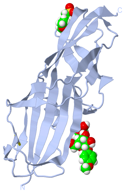
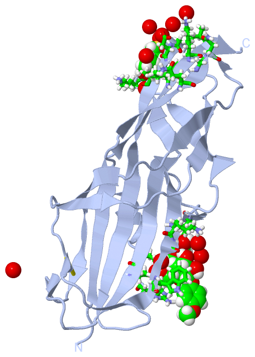
Chain A from PDB  Type:PROTEIN  Length:198
                                                                                                                                                                                                                                      
               SCOP domains ------------------------------------------------------------------------------------------------------------------------------------------------------------------------------------------------------ SCOP domains
               CATH domains ------------------------------------------------------------------------------------------------------------------------------------------------------------------------------------------------------ CATH domains
               Pfam domains ------------------------------------------------------------------------------------------------------------------------------------------------------------------------------------------------------ Pfam domains
         Sec.struct. author ...eeeeeeee..eeeeeeeeee.....eeee....eeeeeeeeee....ee.....eeeeeeeeeeee.eee.....eeeeeeee....eee......eeeeeeeeeeeeee...ee.......eeee......eeeeee.......eeeeeeeeeeeeeeeeee.........hhhhhh.....eeeeeeeeeee. Sec.struct. author
                 SAPs(SNPs) ------------------------------------------------------------------------------------------------------------------------------------------------------------------------------------------------------ SAPs(SNPs)
                    PROSITE ------------------------------------------------------------------------------------------------------------------------------------------------------------------------------------------------------ PROSITE
                 Transcript ------------------------------------------------------------------------------------------------------------------------------------------------------------------------------------------------------ Transcript
                 4z3g A  -1 MAWNNIVFYSLGDVNSYQGGNVVITQRPQFITSWRPGIATVTWNQCNGPEFADGSWAYYREYIAWVVFPKKVMTKNGYPLFIEVHNKGSWSEENTGDNDSYFFLKGYKWDQRAFDTANLCQKPGETTRLTEKFDDIIFKVALPADLPLGDYSVTIPYTSGIQRHFASYLGARFKIPYNVAKTLPRENEMLFLFKNIGG 196
                                     8        18        28        38        48        58        68        78        88        98       108       118       128       138       148       158       168       178       188        

Chain B from PDB  Type:PROTEIN  Length:198
                                                                                                                                                                                                                                      
               SCOP domains ------------------------------------------------------------------------------------------------------------------------------------------------------------------------------------------------------ SCOP domains
               CATH domains ------------------------------------------------------------------------------------------------------------------------------------------------------------------------------------------------------ CATH domains
               Pfam domains ------------------------------------------------------------------------------------------------------------------------------------------------------------------------------------------------------ Pfam domains
         Sec.struct. author ...eeeeeeee..eeeeeeeeee.....eeee....eeeeeeeeee....ee.....eeeeeeeeeeee.eee.....eeeeeeee....eee......eeeeeeeeeeeeee...ee.......eeee......eeeeee.......eeeeeeeeeeeeeeeeee.........hhhhhh.....eeeeeeeeeee. Sec.struct. author
                 SAPs(SNPs) ------------------------------------------------------------------------------------------------------------------------------------------------------------------------------------------------------ SAPs(SNPs)
                    PROSITE ------------------------------------------------------------------------------------------------------------------------------------------------------------------------------------------------------ PROSITE
                 Transcript ------------------------------------------------------------------------------------------------------------------------------------------------------------------------------------------------------ Transcript
                 4z3g B  -1 MAWNNIVFYSLGDVNSYQGGNVVITQRPQFITSWRPGIATVTWNQCNGPEFADGSWAYYREYIAWVVFPKKVMTKNGYPLFIEVHNKGSWSEENTGDNDSYFFLKGYKWDQRAFDTANLCQKPGETTRLTEKFDDIIFKVALPADLPLGDYSVTIPYTSGIQRHFASYLGARFKIPYNVAKTLPRENEMLFLFKNIGG 196
                                     8        18        28        38        48        58        68        78        88        98       108       118       128       138       148       158       168       178       188        

   Legend:   → Mismatch (orange background)
  - → Gap (green background, '-', border residues have a numbering label)
    → Modified Residue (blue background, lower-case, 'x' indicates undefined single-letter code, labelled with number + name)
  x → Chemical Group (purple background, 'x', labelled with number + name, e.g. ACE or NH2)
  extra numbering lines below/above indicate numbering irregularities and modified residue names etc., number ends below/above '|'

 Classification and Annotation

(-) SCOP Domains  (0, 0)

(no "SCOP Domain" information available for 4Z3G)

(-) CATH Domains  (0, 0)

(no "CATH Domain" information available for 4Z3G)

(-) Pfam Domains  (0, 0)

(no "Pfam Domain" information available for 4Z3G)

(-) Gene Ontology  (0, 0)

Asymmetric Unit(hide GO term definitions)
    (no "Gene Ontology" information available for 4Z3G)

 Visualization

(-) Interactive Views

Asymmetric Unit
  Complete Structure
    Jena3D(integrated viewing of ligand, site, SAP, PROSITE, SCOP information)
    WebMol | AstexViewer[tm]@PDBe
(Java Applets, require no local installation except for Java; loading may be slow)
    STRAP
(Java WebStart application, automatic local installation, requires Java; full application with system access!)
    RasMol
(require local installation)
    Molscript (VRML)
(requires installation of a VRML viewer; select preferred view via VRML and generate a mono or stereo PDF format file)
 
  Ligands, Modified Residues, Ions
    4KS  [ RasMol | Jena3D ]  +environment [ RasMol | Jena3D ]
    GAL  [ RasMol | Jena3D ]  +environment [ RasMol | Jena3D ]
    GLA  [ RasMol | Jena3D ]  +environment [ RasMol | Jena3D ]
    MES  [ RasMol | Jena3D ]  +environment [ RasMol | Jena3D ]
 
  Sites
    AC1  [ RasMol ]  +environment [ RasMol ]
    AC2  [ RasMol ]  +environment [ RasMol ]
    AC3  [ RasMol ]  +environment [ RasMol ]
    AC4  [ RasMol ]  +environment [ RasMol ]
 
  Cis Peptide Bonds
(no "Cis Peptide Bonds" information available for 4z3g)
 
Biological Units
  Complete Structure
    Biological Unit 1  [ Jena3D ]
    Biological Unit 2  [ Jena3D ]

(-) Still Images

Jmol
  protein: cartoon or spacefill or dots and stick; nucleic acid: cartoon and stick; ligands: spacefill; active site: stick
Molscript
  protein, nucleic acid: cartoon; ligands: spacefill; active site: ball and stick

 Databases and Analysis Tools

(-) Databases

Access by PDB/NDB ID
  4z3g
    Family and Domain InformationProDom | SYSTERS
    General Structural InformationGlycoscienceDB | MMDB | NDB | OCA | PDB | PDBe | PDBj | PDBsum | PDBWiki | PQS | PROTEOPEDIA
    Orientation in MembranesOPM
    Protein SurfaceSURFACE
    Secondary StructureDSSP (structure derived) | HSSP (homology derived)
    Structural GenomicsGeneCensus
    Structural NeighboursCE | VAST
    Structure ClassificationCATH | Dali | SCOP
    Validation and Original DataBMRB Data View | BMRB Restraints Grid | EDS | PROCHECK | RECOORD | WHAT_CHECK
 
Access by UniProt ID/Accession number
  A0A182DW20_E | A0A182DW20
    Comparative Protein Structure ModelsModBase
    Genomic InformationEnsembl
    Protein-protein InteractionDIP
    Sequence, Family and Domain InformationInterPro | Pfam | SMART | UniProtKB/TrEMBL
 
Access by Enzyme Classificator   (EC Number)
  (no 'Enzyme Classificator' available)
    General Enzyme InformationBRENDA | EC-PDB | Enzyme | IntEnz
    PathwayKEGG | MetaCyc
 
Access by Disease Identifier   (MIM ID)
  (no 'MIM ID' available)
    Disease InformationOMIM
 
Access by GenAge ID
  (no 'GenAge ID' available)
    Age Related InformationGenAge

(-) Analysis Tools

Access by PDB/NDB ID
    Domain InformationXDom
    Interatomic Contacts of Structural UnitsCSU
    Ligand-protein ContactsLPC
    Protein CavitiescastP
    Sequence and Secondary StructurePDBCartoon
    Structure AlignmentSTRAP(Java WebStart application, automatic local installation, requires Java; full application with system access!)
    Structure and Sequence BrowserSTING
 
Access by UniProt ID/Accession number
  A0A182DW20_E | A0A182DW20
    Protein Disorder PredictionDisEMBL | FoldIndex | GLOBPLOT (for more information see DisProt)

 Related Entries

(-) Entries Sharing at Least One Protein Chain (UniProt ID)

UniProtKB/TrEMBL
        A0A182DW20_E | A0A182DW204z3e 4z3f 4z3h 4z3i 4z3j

(-) Related Entries Specified in the PDB File

(no "Related Entries Specified in the PDB File" available for 4Z3G)