Show PDB file:   
         Plain Text   HTML   (compressed file size)
QuickSearch:   
by PDB,NDB,UniProt,PROSITE Code or Search Term(s)  
(-)Asymmetric Unit
(-)Asym. Unit - sites
(-)Biological Unit 1
(-)Biol. Unit 1 - sites
(-)Biological Unit 2
collapse expand < >
Image Asymmetric Unit
Asymmetric Unit  (Jmol Viewer)
Image Asym. Unit - sites
Asym. Unit - sites  (Jmol Viewer)
Image Biological Unit 1
Biological Unit 1  (Jmol Viewer)
Image Biol. Unit 1 - sites
Biol. Unit 1 - sites  (Jmol Viewer)
Image Biological Unit 2
Biological Unit 2  (Jmol Viewer)

(-) Description

Title :  CRYSTAL STRUCTURE OF THE THIRD FNIII DOMAIN OF INTEGRIN BETA4
 
Authors :  N. Alonso-Garcia, H. Urien, J. M. De Pereda
Date :  30 Oct 14  (Deposition) - 11 Feb 15  (Release) - 22 Apr 15  (Revision)
Method :  X-RAY DIFFRACTION
Resolution :  1.61
Chains :  Asym. Unit :  A,B
Biol. Unit 1:  A  (1x)
Biol. Unit 2:  B  (1x)
Keywords :  Immunoglobulin Fold, Fibronectin Type Iii, Integrin, Cell Adhesion (Keyword Search: [Gene Ontology, PubMed, Web (Google))
 
Reference :  N. Alonso-Garcia, I. Garcia-Rubio, J. A. Manso, R. M. Buey, H. Urien, A. Sonnenberg, G. Jeschke, J. M. De Pereda
Combination Of X-Ray Crystallography, Saxs And Deer To Obtain The Structure Of The Fniii-3, 4 Domains Of Integrin Alpha6Beta4
Acta Crystallogr. , Sect. D V. 71 969 2015
PubMed-ID: 25849406  |  Reference-DOI: 10.1107/S1399004715002485

(-) Compounds

Molecule 1 - INTEGRIN BETA-4
    ChainsA, B
    EngineeredYES
    Expression SystemESCHERICHIA COLI BL21(DE3)
    Expression System PlasmidMODIFIED PET15B
    Expression System Taxid469008
    Expression System Vector TypePLASMID
    FragmentTHIRD FNIII DOMAIN, UNP RESIDUES 1457-1548
    GeneITGB4
    Organism CommonHUMAN
    Organism ScientificHOMO SAPIENS
    Organism Taxid9606
    SynonymGP150

 Structural Features

(-) Chains, Units

  12
Asymmetric Unit AB
Biological Unit 1 (1x)A 
Biological Unit 2 (1x) B

Summary Information (see also Sequences/Alignments below)

(-) Ligands, Modified Residues, Ions  (3, 4)

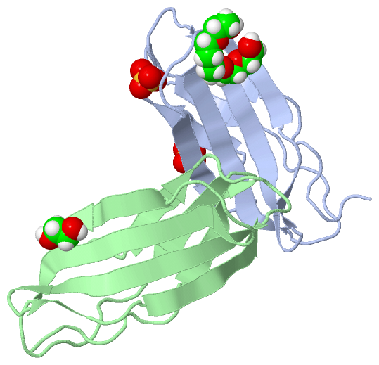
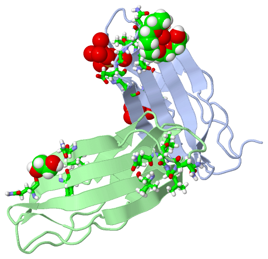
Asymmetric Unit (3, 4)
No.NameCountTypeFull Name
11PE1Ligand/IonPENTAETHYLENE GLYCOL
2EDO1Ligand/Ion1,2-ETHANEDIOL
3SO42Ligand/IonSULFATE ION
Biological Unit 1 (2, 3)
No.NameCountTypeFull Name
11PE1Ligand/IonPENTAETHYLENE GLYCOL
2EDO-1Ligand/Ion1,2-ETHANEDIOL
3SO42Ligand/IonSULFATE ION
Biological Unit 2 (1, 1)
No.NameCountTypeFull Name
11PE-1Ligand/IonPENTAETHYLENE GLYCOL
2EDO1Ligand/Ion1,2-ETHANEDIOL
3SO4-1Ligand/IonSULFATE ION

(-) Sites  (4, 4)

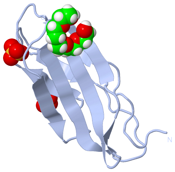
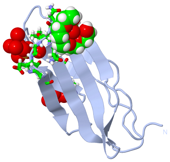
Asymmetric Unit (4, 4)
No.NameEvidenceResiduesDescription
1AC1SOFTWAREGLY A:1500 , GLU A:1501 , HIS A:1503 , HOH A:1715 , HOH A:1764 , HOH A:1772 , HOH A:1778 , HOH A:1804binding site for residue SO4 A 1601
2AC2SOFTWAREARG A:1475 , SER A:1514 , HOH A:1703binding site for residue SO4 A 1602
3AC3SOFTWAREGLN A:1495 , LEU A:1496 , LEU A:1497 , ASN A:1498 , GLY A:1499 , HOH A:1727 , GLN B:1495 , LEU B:1496 , LEU B:1497 , ASN B:1498 , GLY B:1499 , VAL B:1527binding site for residue 1PE A 1603
4AC4SOFTWAREGLN B:1488 , TYR B:1490 , ASN B:1506 , ILE B:1507binding site for residue EDO B 1601

(-) SS Bonds  (0, 0)

(no "SS Bond" information available for 4WTW)

(-) Cis Peptide Bonds  (0, 0)

(no "Cis Peptide Bond" information available for 4WTW)

 Sequence-Structure Mapping

(-) SAPs(SNPs)/Variants  (0, 0)

(no "SAP(SNP)/Variant" information available for 4WTW)

(-) PROSITE Motifs  (0, 0)

(no "PROSITE Motif" information available for 4WTW)

(-) Exons   (0, 0)

(no "Exon" information available for 4WTW)

(-) Sequences/Alignments

Asymmetric Unit
   Reformat: Number of residues per line =  ('0' or empty: single-line sequence representation)
  Number of residues per labelling interval =   
  UniProt sequence: complete  aligned part    
   Show mapping: SCOP domains CATH domains Pfam domains Secondary structure (by author)
SAPs(SNPs) PROSITE motifs Exons
(details for a mapped element are shown in a popup box when the mouse pointer rests over it)
Chain A from PDB  Type:PROTEIN  Length:95
                                                                                                                                
               SCOP domains ----------------------------------------------------------------------------------------------- SCOP domains
               CATH domains ----------------------------------------------------------------------------------------------- CATH domains
               Pfam domains ----------------------------------------------------------------------------------------------- Pfam domains
         Sec.struct. author ...........eeee.....eeee.........eeeeeeeeee......eeeee......eeee.......eeeeeeeeee..ee...eeeeee. Sec.struct. author
                 SAPs(SNPs) ----------------------------------------------------------------------------------------------- SAPs(SNPs)
                    PROSITE ----------------------------------------------------------------------------------------------- PROSITE
                 Transcript ----------------------------------------------------------------------------------------------- Transcript
                4wtw A 1454 SHMVPDTPTRLVFSALGPTSLRVSWQEPRCERPLQGYSVEYQLLNGGELHRLNIPNPAQTSVVVEDLLPNHSYVFRVRAQSQEGWGREREGVITI 1548
                                  1463      1473      1483      1493      1503      1513      1523      1533      1543     

Chain B from PDB  Type:PROTEIN  Length:93
                                                                                                                              
               SCOP domains --------------------------------------------------------------------------------------------- SCOP domains
               CATH domains --------------------------------------------------------------------------------------------- CATH domains
               Pfam domains --------------------------------------------------------------------------------------------- Pfam domains
         Sec.struct. author .........eeeeee..eeeee.........eeeeeeeeee......eeeee......eeee.......eeeeeeeeee..ee...eeeeee. Sec.struct. author
                 SAPs(SNPs) --------------------------------------------------------------------------------------------- SAPs(SNPs)
                    PROSITE --------------------------------------------------------------------------------------------- PROSITE
                 Transcript --------------------------------------------------------------------------------------------- Transcript
                4wtw B 1456 MVPDTPTRLVFSALGPTSLRVSWQEPRCERPLQGYSVEYQLLNGGELHRLNIPNPAQTSVVVEDLLPNHSYVFRVRAQSQEGWGREREGVITI 1548
                                  1465      1475      1485      1495      1505      1515      1525      1535      1545   

   Legend:   → Mismatch (orange background)
  - → Gap (green background, '-', border residues have a numbering label)
    → Modified Residue (blue background, lower-case, 'x' indicates undefined single-letter code, labelled with number + name)
  x → Chemical Group (purple background, 'x', labelled with number + name, e.g. ACE or NH2)
  extra numbering lines below/above indicate numbering irregularities and modified residue names etc., number ends below/above '|'

 Classification and Annotation

(-) SCOP Domains  (0, 0)

(no "SCOP Domain" information available for 4WTW)

(-) CATH Domains  (0, 0)

(no "CATH Domain" information available for 4WTW)

(-) Pfam Domains  (0, 0)

(no "Pfam Domain" information available for 4WTW)

(-) Gene Ontology  (31, 31)

Asymmetric Unit(hide GO term definitions)

 Visualization

(-) Interactive Views

Asymmetric Unit
  Complete Structure
    Jena3D(integrated viewing of ligand, site, SAP, PROSITE, SCOP information)
    WebMol | AstexViewer[tm]@PDBe
(Java Applets, require no local installation except for Java; loading may be slow)
    STRAP
(Java WebStart application, automatic local installation, requires Java; full application with system access!)
    RasMol
(require local installation)
    Molscript (VRML)
(requires installation of a VRML viewer; select preferred view via VRML and generate a mono or stereo PDF format file)
 
  Ligands, Modified Residues, Ions
    1PE  [ RasMol | Jena3D ]  +environment [ RasMol | Jena3D ]
    EDO  [ RasMol | Jena3D ]  +environment [ RasMol | Jena3D ]
    SO4  [ RasMol | Jena3D ]  +environment [ RasMol | Jena3D ]
 
  Sites
    AC1  [ RasMol ]  +environment [ RasMol ]
    AC2  [ RasMol ]  +environment [ RasMol ]
    AC3  [ RasMol ]  +environment [ RasMol ]
    AC4  [ RasMol ]  +environment [ RasMol ]
 
  Cis Peptide Bonds
(no "Cis Peptide Bonds" information available for 4wtw)
 
Biological Units
  Complete Structure
    Biological Unit 1  [ Jena3D ]
    Biological Unit 2  [ Jena3D ]

(-) Still Images

Jmol
  protein: cartoon or spacefill or dots and stick; nucleic acid: cartoon and stick; ligands: spacefill; active site: stick
Molscript
  protein, nucleic acid: cartoon; ligands: spacefill; active site: ball and stick

 Databases and Analysis Tools

(-) Databases

Access by PDB/NDB ID
  4wtw
    Family and Domain InformationProDom | SYSTERS
    General Structural InformationGlycoscienceDB | MMDB | NDB | OCA | PDB | PDBe | PDBj | PDBsum | PDBWiki | PQS | PROTEOPEDIA
    Orientation in MembranesOPM
    Protein SurfaceSURFACE
    Secondary StructureDSSP (structure derived) | HSSP (homology derived)
    Structural GenomicsGeneCensus
    Structural NeighboursCE | VAST
    Structure ClassificationCATH | Dali | SCOP
    Validation and Original DataBMRB Data View | BMRB Restraints Grid | EDS | PROCHECK | RECOORD | WHAT_CHECK
 
Access by UniProt ID/Accession number
  ITB4_HUMAN | P16144
    Comparative Protein Structure ModelsModBase
    Genomic InformationEnsembl
    Protein-protein InteractionDIP
    Sequence, Family and Domain InformationInterPro | Pfam | SMART | UniProtKB/SwissProt
 
Access by Enzyme Classificator   (EC Number)
  (no 'Enzyme Classificator' available)
    General Enzyme InformationBRENDA | EC-PDB | Enzyme | IntEnz
    PathwayKEGG | MetaCyc
 
Access by Disease Identifier   (MIM ID)
  (no 'MIM ID' available)
    Disease InformationOMIM
 
Access by GenAge ID
  (no 'GenAge ID' available)
    Age Related InformationGenAge

(-) Analysis Tools

Access by PDB/NDB ID
    Domain InformationXDom
    Interatomic Contacts of Structural UnitsCSU
    Ligand-protein ContactsLPC
    Protein CavitiescastP
    Sequence and Secondary StructurePDBCartoon
    Structure AlignmentSTRAP(Java WebStart application, automatic local installation, requires Java; full application with system access!)
    Structure and Sequence BrowserSTING
 
Access by UniProt ID/Accession number
  ITB4_HUMAN | P16144
    Protein Disorder PredictionDisEMBL | FoldIndex | GLOBPLOT (for more information see DisProt)

 Related Entries

(-) Entries Sharing at Least One Protein Chain (UniProt ID)

UniProtKB/Swiss-Prot
        ITB4_HUMAN | P161441qg3 2yrz 3f7p 3f7q 3f7r 3fq4 3fso 3h6a 4q58 4wtx

(-) Related Entries Specified in the PDB File

2yrz NMR STRUCTURE OF THIS DOMAIN OF INTEGRIN BETA4. NOT IDENTICAL CONSTRUCT.