Show PDB file:   
         Plain Text   HTML   (compressed file size)
QuickSearch:   
by PDB,NDB,UniProt,PROSITE Code or Search Term(s)  
(-)Asym./Biol. Unit
(-)Asym./Biol. Unit - sites
collapse expand < >
Image Asym./Biol. Unit
Asym./Biol. Unit  (Jmol Viewer)
Image Asym./Biol. Unit - sites
Asym./Biol. Unit - sites  (Jmol Viewer)

(-) Description

Title :  CRYSTAL STRUCTURE OF NS3/4A PROTEASE IN COMPLEX WITH ASUNAPREVIR
 
Authors :  C. A. Schiffer, D. I. Soumana, A. Ali
Date :  13 Sep 14  (Deposition) - 08 Oct 14  (Release) - 25 Nov 15  (Revision)
Method :  X-RAY DIFFRACTION
Resolution :  1.70
Chains :  Asym./Biol. Unit :  A
Keywords :  Hcv Protease Inhibitor Complex, Resistance, Hydrolase-Hydrolase Inhibitor Complex (Keyword Search: [Gene Ontology, PubMed, Web (Google))
 
Reference :  D. I. Soumana, A. Ali, C. A. Schiffer
Structural Analysis Of Asunaprevir Resistance In Hcv Ns3/4A Protease.
Acs Chem. Biol. V. 9 2485 2014
PubMed-ID: 25243902  |  Reference-DOI: 10.1021/CB5006118

(-) Compounds

Molecule 1 - NS3 PROTEIN
    ChainsA
    EngineeredYES
    Expression SystemESCHERICHIA COLI
    Expression System PlasmidPET28A
    Expression System StrainBL21 (DE3)
    Expression System Taxid469008
    Organism ScientificHEPATITIS C VIRUS
    Organism Taxid11103

 Structural Features

(-) Chains, Units

  1
Asymmetric/Biological Unit A

Summary Information (see also Sequences/Alignments below)

(-) Ligands, Modified Residues, Ions  (3, 3)

Asymmetric/Biological Unit (3, 3)
No.NameCountTypeFull Name
12R91Ligand/IonN-(TERT-BUTOXYCARBONYL)-3-METHYL-L-VALYL-(4R)-4-[(7-CHLORO-4-METHOXYISOQUINOLIN-1-YL)OXY]-N-{(1R,2S)-1-[(CYCLOPROPYLSULFONYL)CARBAMOYL]-2-ETHENYLCYCLOPROPYL}-L-PROLINAMIDE
2CL1Ligand/IonCHLORIDE ION
3ZN1Ligand/IonZINC ION

(-) Sites  (3, 3)

Asymmetric Unit (3, 3)
No.NameEvidenceResiduesDescription
1AC1SOFTWARECYS A:1097 , CYS A:1099 , CYS A:1145 , HIS A:1149binding site for residue ZN A 1201
2AC2SOFTWAREILE A:994 , ARG A:1011 , ASP A:1025 , ASN A:1027 , HOH A:1350binding site for residue CL A 1202
3AC3SOFTWAREARG A:997 , TYR A:1006 , ALA A:1007 , GLN A:1008 , GLU A:1030 , PHE A:1043 , HIS A:1057 , VAL A:1078 , ASP A:1081 , ARG A:1123 , ILE A:1132 , LEU A:1135 , LYS A:1136 , GLY A:1137 , SER A:1139 , PHE A:1154 , ARG A:1155 , ALA A:1156 , ALA A:1157 , ASP A:1168 , HOH A:1363 , HOH A:1385 , HOH A:1394binding site for residue 2R9 A 1203

(-) SS Bonds  (0, 0)

(no "SS Bond" information available for 4WF8)

(-) Cis Peptide Bonds  (1, 1)

Asymmetric/Biological Unit
No.Residues
1Ala A:1014 -Ala A:1015

 Sequence-Structure Mapping

(-) SAPs(SNPs)/Variants  (0, 0)

(no "SAP(SNP)/Variant" information available for 4WF8)

(-) PROSITE Motifs  (0, 0)

(no "PROSITE Motif" information available for 4WF8)

(-) Exons   (0, 0)

(no "Exon" information available for 4WF8)

(-) Sequences/Alignments

Asymmetric/Biological Unit
   Reformat: Number of residues per line =  ('0' or empty: single-line sequence representation)
  Number of residues per labelling interval =   
  UniProt sequence: complete  aligned part    
   Show mapping: SCOP domains CATH domains Pfam domains Secondary structure (by author)
SAPs(SNPs) PROSITE motifs Exons
(details for a mapped element are shown in a popup box when the mouse pointer rests over it)
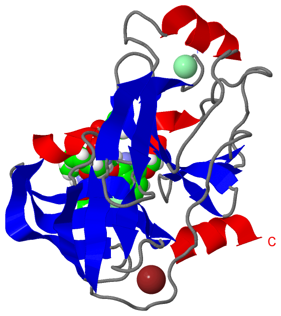
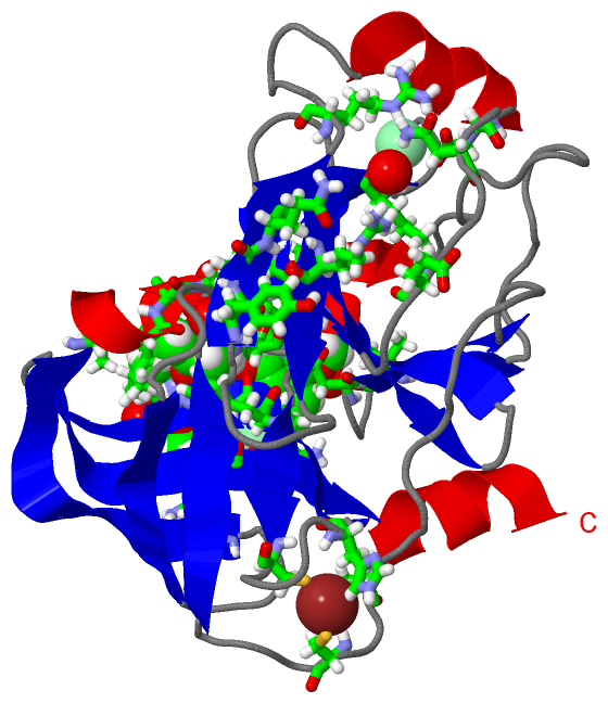
Chain A from PDB  Type:PROTEIN  Length:192
                                                                                                                                                                                                                                 
               SCOP domains ------------------------------------------------------------------------------------------------------------------------------------------------------------------------------------------------ SCOP domains
               CATH domains ------------------------------------------------------------------------------------------------------------------------------------------------------------------------------------------------ CATH domains
               Pfam domains ------------------------------------------------------------------------------------------------------------------------------------------------------------------------------------------------ Pfam domains
         Sec.struct. author ....eeeeeee......eeee......hhhhhhhh.........eeeee....eeeeeee..eeeeehhhhh..............eee....eeeee................eeeee.....eeeeee....eeeeeeeeehhhhh.....eee.....eeeeeeeeeee..eeeeeeeeehhhhhhhhh Sec.struct. author
                 SAPs(SNPs) ------------------------------------------------------------------------------------------------------------------------------------------------------------------------------------------------ SAPs(SNPs)
                    PROSITE ------------------------------------------------------------------------------------------------------------------------------------------------------------------------------------------------ PROSITE
                 Transcript ------------------------------------------------------------------------------------------------------------------------------------------------------------------------------------------------ Transcript
                4wf8 A  989 KGSVVIVGRINLSGDTAYAQQTRGAAACQETSQTGRDKNQVEGEVQIVSTATQTFLATSINGVLWTVYHGAGTRTIASPKGPVTQMYTNVDKDLVGWQAPQGSRSLTPCTCGSSDLYLVTRHADVIPVRRRGDSRGSLLSPRPISYLKGSSGGPLLCPAGHAVGIFRAAVSTRGVAKAVDFIPVESLETTMR 1180
                                   998      1008      1018      1028      1038      1048      1058      1068      1078      1088      1098      1108      1118      1128      1138      1148      1158      1168      1178  

   Legend:   → Mismatch (orange background)
  - → Gap (green background, '-', border residues have a numbering label)
    → Modified Residue (blue background, lower-case, 'x' indicates undefined single-letter code, labelled with number + name)
  x → Chemical Group (purple background, 'x', labelled with number + name, e.g. ACE or NH2)
  extra numbering lines below/above indicate numbering irregularities and modified residue names etc., number ends below/above '|'

 Classification and Annotation

(-) SCOP Domains  (0, 0)

(no "SCOP Domain" information available for 4WF8)

(-) CATH Domains  (0, 0)

(no "CATH Domain" information available for 4WF8)

(-) Pfam Domains  (0, 0)

(no "Pfam Domain" information available for 4WF8)

(-) Gene Ontology  (4, 4)

Asymmetric/Biological Unit(hide GO term definitions)

 Visualization

(-) Interactive Views

Asymmetric/Biological Unit
  Complete Structure
    Jena3D(integrated viewing of ligand, site, SAP, PROSITE, SCOP information)
    WebMol | AstexViewer[tm]@PDBe
(Java Applets, require no local installation except for Java; loading may be slow)
    STRAP
(Java WebStart application, automatic local installation, requires Java; full application with system access!)
    RasMol
(require local installation)
    Molscript (VRML)
(requires installation of a VRML viewer; select preferred view via VRML and generate a mono or stereo PDF format file)
 
  Ligands, Modified Residues, Ions
    2R9  [ RasMol | Jena3D ]  +environment [ RasMol | Jena3D ]
    CL  [ RasMol | Jena3D ]  +environment [ RasMol | Jena3D ]
    ZN  [ RasMol | Jena3D ]  +environment [ RasMol | Jena3D ]
 
  Sites
    AC1  [ RasMol ]  +environment [ RasMol ]
    AC2  [ RasMol ]  +environment [ RasMol ]
    AC3  [ RasMol ]  +environment [ RasMol ]
 
  Cis Peptide Bonds
    Ala A:1014 - Ala A:1015   [ RasMol ]  
 

(-) Still Images

Jmol
  protein: cartoon or spacefill or dots and stick; nucleic acid: cartoon and stick; ligands: spacefill; active site: stick
Molscript
  protein, nucleic acid: cartoon; ligands: spacefill; active site: ball and stick

 Databases and Analysis Tools

(-) Databases

Access by PDB/NDB ID
  4wf8
    Family and Domain InformationProDom | SYSTERS
    General Structural InformationGlycoscienceDB | MMDB | NDB | OCA | PDB | PDBe | PDBj | PDBsum | PDBWiki | PQS | PROTEOPEDIA
    Orientation in MembranesOPM
    Protein SurfaceSURFACE
    Secondary StructureDSSP (structure derived) | HSSP (homology derived)
    Structural GenomicsGeneCensus
    Structural NeighboursCE | VAST
    Structure ClassificationCATH | Dali | SCOP
    Validation and Original DataBMRB Data View | BMRB Restraints Grid | EDS | PROCHECK | RECOORD | WHAT_CHECK
 
Access by UniProt ID/Accession number
  X2G809_9HEPC | X2G809
    Comparative Protein Structure ModelsModBase
    Genomic InformationEnsembl
    Protein-protein InteractionDIP
    Sequence, Family and Domain InformationInterPro | Pfam | SMART | UniProtKB/TrEMBL
 
Access by Enzyme Classificator   (EC Number)
  (no 'Enzyme Classificator' available)
    General Enzyme InformationBRENDA | EC-PDB | Enzyme | IntEnz
    PathwayKEGG | MetaCyc
 
Access by Disease Identifier   (MIM ID)
  (no 'MIM ID' available)
    Disease InformationOMIM
 
Access by GenAge ID
  (no 'GenAge ID' available)
    Age Related InformationGenAge

(-) Analysis Tools

Access by PDB/NDB ID
    Domain InformationXDom
    Interatomic Contacts of Structural UnitsCSU
    Ligand-protein ContactsLPC
    Protein CavitiescastP
    Sequence and Secondary StructurePDBCartoon
    Structure AlignmentSTRAP(Java WebStart application, automatic local installation, requires Java; full application with system access!)
    Structure and Sequence BrowserSTING
 
Access by UniProt ID/Accession number
  X2G809_9HEPC | X2G809
    Protein Disorder PredictionDisEMBL | FoldIndex | GLOBPLOT (for more information see DisProt)

 Related Entries

(-) Entries Sharing at Least One Protein Chain (UniProt ID)

(no "Entries Sharing at Least One Protein Chain" available for 4WF8)

(-) Related Entries Specified in the PDB File

(no "Related Entries Specified in the PDB File" available for 4WF8)