Show PDB file:   
         Plain Text   HTML   (compressed file size)
QuickSearch:   
by PDB,NDB,UniProt,PROSITE Code or Search Term(s)  
(-)Asymmetric Unit
(-)Asym. Unit - sites
(-)Biological Unit 1
(-)Biol. Unit 1 - sites
collapse expand < >
Image Asymmetric Unit
Asymmetric Unit  (Jmol Viewer)
Image Asym. Unit - sites
Asym. Unit - sites  (Jmol Viewer)
Image Biological Unit 1
Biological Unit 1  (Jmol Viewer)
Image Biol. Unit 1 - sites
Biol. Unit 1 - sites  (Jmol Viewer)

(-) Description

Title :  CRYSTAL STRUCTURE OF THE ETS DOMAIN OF HUMAN ETV5 IN COMPLEX WITH DNA
 
Authors :  J. A. Newman, H. Aitkenhead, C. D. O. Cooper, D. M. Pinkas, L. Shrestha, N. Burgess-Brown, J. Kopec, F. Fitzpatrick, C. Tallant, F. Von Delft C. H. Arrowsmith, C. Bountra, A. Edwards, O. Gileadi
Date :  29 May 14  (Deposition) - 11 Jun 14  (Release) - 10 Jun 15  (Revision)
Method :  X-RAY DIFFRACTION
Resolution :  1.95
Chains :  Asym. Unit :  A,B,C
Biol. Unit 1:  A,B,C  (2x)
Keywords :  Dna Binding Protein (Keyword Search: [Gene Ontology, PubMed, Web (Google))
 
Reference :  C. D. O. Cooper, J. A. Newman, H. Aitkenhead, C. K. Allerston, O. Gileadi
Structures Of The Ets Domains Of Transcription Factors Etv1 Etv4, Etv5 And Fev: Determinants Of Dna Binding And Redox Regulation By Disulfide Bond Formation.
J. Biol. Chem. V. 290 13692 2015
PubMed-ID: 25866208  |  Reference-DOI: 10.1074/JBC.M115.646737

(-) Compounds

Molecule 1 - ETS TRANSLOCATION VARIANT 5
    ChainsA
    EngineeredYES
    Expression SystemESCHERICHIA COLI
    Expression System StrainBL21(DE3)
    Expression System Taxid469008
    FragmentETS DOMAIN, RESIDUES 365-462
    Organism CommonHUMAN
    Organism ScientificHOMO SAPIENS
    Organism Taxid9606
    SynonymETS-RELATED PROTEIN ERM
 
Molecule 2 - 5'-D(*AP*CP*CP*GP*GP*AP*AP*GP*TP*GP)-3'
    ChainsB
    Organism ScientificSYNTHETIC CONSTRUCT
    Organism Taxid32630
    SyntheticYES
 
Molecule 3 - 5'-D(*AP*CP*TP*TP*CP*CP*GP*GP*TP*CP)-3'
    ChainsC
    Organism ScientificSYNTHETIC CONSTRUCT
    Organism Taxid32630
    SyntheticYES

 Structural Features

(-) Chains, Units

  123
Asymmetric Unit ABC
Biological Unit 1 (2x)ABC

Summary Information (see also Sequences/Alignments below)

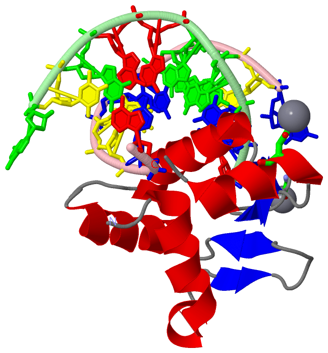
(-) Ligands, Modified Residues, Ions  (1, 2)

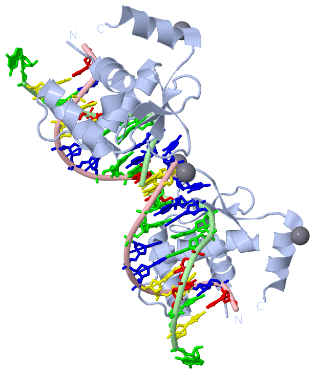
Asymmetric Unit (1, 2)
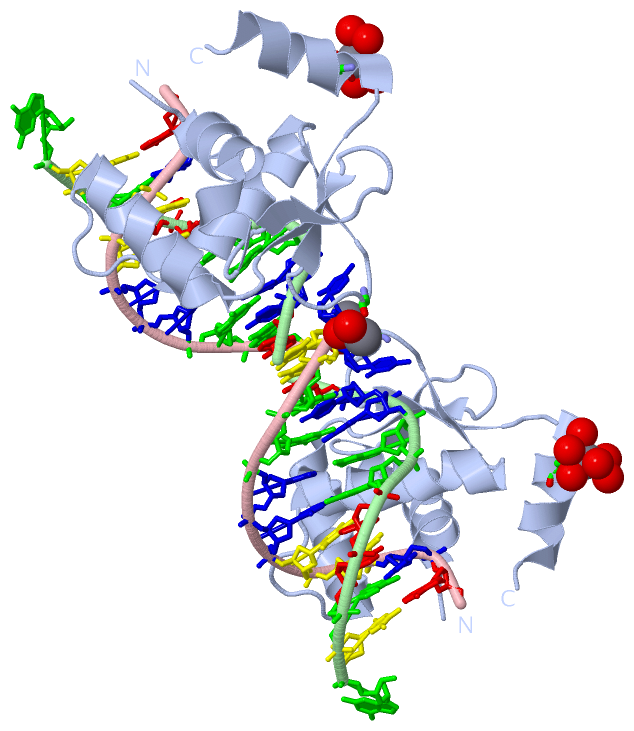
No.NameCountTypeFull Name
1CA2Ligand/IonCALCIUM ION
Biological Unit 1 (0, 0)
No.NameCountTypeFull Name
1CA-1Ligand/IonCALCIUM ION

(-) Sites  (2, 2)

Asymmetric Unit (2, 2)
No.NameEvidenceResiduesDescription
1AC1SOFTWAREGLY A:440 , HOH A:2061 , HOH A:2062BINDING SITE FOR RESIDUE CA A1462
2AC2SOFTWAREASP A:452 , HOH A:2070 , HOH A:2071 , HOH A:2072 , HOH A:2074 , HOH A:2075 , HOH A:2077BINDING SITE FOR RESIDUE CA A1463

(-) SS Bonds  (0, 0)

(no "SS Bond" information available for 4UNO)

(-) Cis Peptide Bonds  (0, 0)

(no "Cis Peptide Bond" information available for 4UNO)

 Sequence-Structure Mapping

(-) SAPs(SNPs)/Variants  (0, 0)

(no "SAP(SNP)/Variant" information available for 4UNO)

(-) PROSITE Motifs  (0, 0)

(no "PROSITE Motif" information available for 4UNO)

(-) Exons   (0, 0)

(no "Exon" information available for 4UNO)

(-) Sequences/Alignments

Asymmetric Unit
   Reformat: Number of residues per line =  ('0' or empty: single-line sequence representation)
  Number of residues per labelling interval =   
  UniProt sequence: complete  aligned part    
   Show mapping: SCOP domains CATH domains Pfam domains Secondary structure (by author)
SAPs(SNPs) PROSITE motifs Exons
(details for a mapped element are shown in a popup box when the mouse pointer rests over it)
Chain A from PDB  Type:PROTEIN  Length:95
                                                                                                                               
               SCOP domains ----------------------------------------------------------------------------------------------- SCOP domains
               CATH domains ----------------------------------------------------------------------------------------------- CATH domains
               Pfam domains ----------------------------------------------------------------------------------------------- Pfam domains
         Sec.struct. author ..hhhhhhhhhhhhhhhh...ee.....eee..hhhhhhhhhhhhh.....hhhhhhhhhhhhhhh..eee......eeee..hhhhhhhhhh.. Sec.struct. author
                 SAPs(SNPs) ----------------------------------------------------------------------------------------------- SAPs(SNPs)
                    PROSITE ----------------------------------------------------------------------------------------------- PROSITE
                 Transcript ----------------------------------------------------------------------------------------------- Transcript
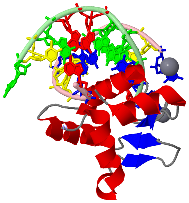
                 4uno A 367 SLQLWQFLVTLLDDPANAHFIAWTGRGMEFKLIEPEEVARRWGIQKNRPAMNYDKLSRSLRYYYEKGIMQKVAGERYVYKFVCDPDALFSMAFPD 461
                                   376       386       396       406       416       426       436       446       456     

Chain B from PDB  Type:DNA  Length:10
                                          
                 4uno B   1 ACCGGAAGTG  10
                                    10

Chain C from PDB  Type:DNA  Length:10
                                          
                 4uno C   1 ACTTCCGGTC  10
                                    10

   Legend:   → Mismatch (orange background)
  - → Gap (green background, '-', border residues have a numbering label)
    → Modified Residue (blue background, lower-case, 'x' indicates undefined single-letter code, labelled with number + name)
  x → Chemical Group (purple background, 'x', labelled with number + name, e.g. ACE or NH2)
  extra numbering lines below/above indicate numbering irregularities and modified residue names etc., number ends below/above '|'

 Classification and Annotation

(-) SCOP Domains  (0, 0)

(no "SCOP Domain" information available for 4UNO)

(-) CATH Domains  (0, 0)

(no "CATH Domain" information available for 4UNO)

(-) Pfam Domains  (0, 0)

(no "Pfam Domain" information available for 4UNO)

(-) Gene Ontology  (24, 24)

Asymmetric Unit(hide GO term definitions)

 Visualization

(-) Interactive Views

Asymmetric Unit
  Complete Structure
    Jena3D(integrated viewing of ligand, site, SAP, PROSITE, SCOP information)
    WebMol | AstexViewer[tm]@PDBe
(Java Applets, require no local installation except for Java; loading may be slow)
    STRAP
(Java WebStart application, automatic local installation, requires Java; full application with system access!)
    RasMol
(require local installation)
    Molscript (VRML)
(requires installation of a VRML viewer; select preferred view via VRML and generate a mono or stereo PDF format file)
 
  Ligands, Modified Residues, Ions
    CA  [ RasMol | Jena3D ]  +environment [ RasMol | Jena3D ]
 
  Sites
    AC1  [ RasMol ]  +environment [ RasMol ]
    AC2  [ RasMol ]  +environment [ RasMol ]
 
  Cis Peptide Bonds
(no "Cis Peptide Bonds" information available for 4uno)
 
Biological Unit
  Complete Structure
    Biological Unit 1  [ Jena3D ]

(-) Still Images

Jmol
  protein: cartoon or spacefill or dots and stick; nucleic acid: cartoon and stick; ligands: spacefill; active site: stick
Molscript
  protein, nucleic acid: cartoon; ligands: spacefill; active site: ball and stick

 Databases and Analysis Tools

(-) Databases

Access by PDB/NDB ID
  4uno
    Family and Domain InformationProDom | SYSTERS
    General Structural InformationGlycoscienceDB | MMDB | NDB | OCA | PDB | PDBe | PDBj | PDBsum | PDBWiki | PQS | PROTEOPEDIA
    Orientation in MembranesOPM
    Protein SurfaceSURFACE
    Secondary StructureDSSP (structure derived) | HSSP (homology derived)
    Structural GenomicsGeneCensus
    Structural NeighboursCE | VAST
    Structure ClassificationCATH | Dali | SCOP
    Validation and Original DataBMRB Data View | BMRB Restraints Grid | EDS | PROCHECK | RECOORD | WHAT_CHECK
 
Access by UniProt ID/Accession number
  ETV5_HUMAN | P41161
    Comparative Protein Structure ModelsModBase
    Genomic InformationEnsembl
    Protein-protein InteractionDIP
    Sequence, Family and Domain InformationInterPro | Pfam | SMART | UniProtKB/SwissProt
 
Access by Enzyme Classificator   (EC Number)
  (no 'Enzyme Classificator' available)
    General Enzyme InformationBRENDA | EC-PDB | Enzyme | IntEnz
    PathwayKEGG | MetaCyc
 
Access by Disease Identifier   (MIM ID)
  (no 'MIM ID' available)
    Disease InformationOMIM
 
Access by GenAge ID
  (no 'GenAge ID' available)
    Age Related InformationGenAge

(-) Analysis Tools

Access by PDB/NDB ID
    Domain InformationXDom
    Interatomic Contacts of Structural UnitsCSU
    Ligand-protein ContactsLPC
    Protein CavitiescastP
    Sequence and Secondary StructurePDBCartoon
    Structure AlignmentSTRAP(Java WebStart application, automatic local installation, requires Java; full application with system access!)
    Structure and Sequence BrowserSTING
 
Access by UniProt ID/Accession number
  ETV5_HUMAN | P41161
    Protein Disorder PredictionDisEMBL | FoldIndex | GLOBPLOT (for more information see DisProt)

 Related Entries

(-) Entries Sharing at Least One Protein Chain (UniProt ID)

UniProtKB/Swiss-Prot
        ETV5_HUMAN | P411615ilv

(-) Related Entries Specified in the PDB File

(no "Related Entries Specified in the PDB File" available for 4UNO)