Show PDB file:   
         Plain Text   HTML   (compressed file size)
QuickSearch:   
by PDB,NDB,UniProt,PROSITE Code or Search Term(s)  
(-)Asymmetric Unit
(-)Asym. Unit - sites
(-)Biological Unit 1
(-)Biol. Unit 1 - sites
(-)Biological Unit 2
(-)Biological Unit 3
collapse expand < >
Image Asymmetric Unit
Asymmetric Unit  (Jmol Viewer)
Image Asym. Unit - sites
Asym. Unit - sites  (Jmol Viewer)
Image Biological Unit 1
Biological Unit 1  (Jmol Viewer)
Image Biol. Unit 1 - sites
Biol. Unit 1 - sites  (Jmol Viewer)
Image Biological Unit 2
Biological Unit 2  (Jmol Viewer)
Image Biological Unit 3
Biological Unit 3  (Jmol Viewer)

(-) Description

Title :  CRYSTAL STRUCTURE OF HUMAN ODORANT BINDING PROTEIN OBPIIA
 
Authors :  A. Schiefner, A. Skerra
Date :  21 Nov 14  (Deposition) - 08 Apr 15  (Release) - 10 Jun 15  (Revision)
Method :  X-RAY DIFFRACTION
Resolution :  2.60
Chains :  Asym. Unit :  A,B
Biol. Unit 1:  A  (1x)
Biol. Unit 2:  B  (1x)
Biol. Unit 3:  A,B  (1x)
Keywords :  Human Lipocalin, Lipid Binding Protein, Odorant Binding Protein, Vanillin, Lilial, Fatty Acids, Aliphatic Aldehydes, Nasal Mucus, Transport Protein (Keyword Search: [Gene Ontology, PubMed, Web (Google))
 
Reference :  A. Schiefner, R. Freier, A. Eichinger, A. Skerra
Crystal Structure Of The Human Odorant Binding Protein, Obpiia.
Proteins V. 83 1180 2015
PubMed-ID: 25810031  |  Reference-DOI: 10.1002/PROT.24797

(-) Compounds

Molecule 1 - ODORANT-BINDING PROTEIN 2A
    ChainsA, B
    EngineeredYES
    Expression SystemESCHERICHIA COLI
    Expression System PlasmidPASK75-HIS
    Expression System StrainJM83
    Expression System Taxid562
    Expression System Vector TypePLASMID
    GeneOBP2A
    MutationYES
    Organism CommonHUMAN
    Organism ScientificHOMO SAPIENS
    Organism Taxid9606
    SynonymODORANT-BINDING PROTEIN IIA, OBPIIA

 Structural Features

(-) Chains, Units

  12
Asymmetric Unit AB
Biological Unit 1 (1x)A 
Biological Unit 2 (1x) B
Biological Unit 3 (1x)AB

Summary Information (see also Sequences/Alignments below)

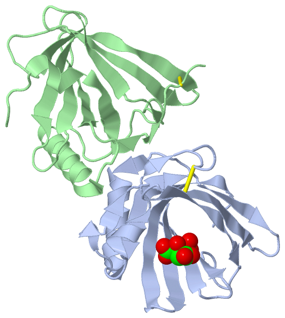
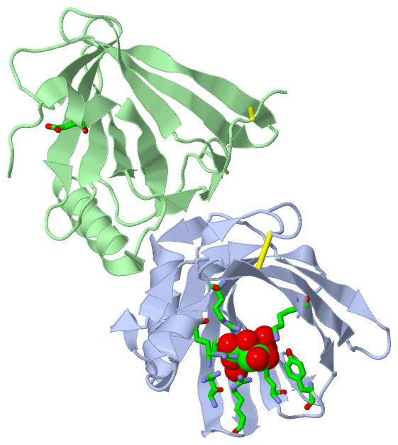
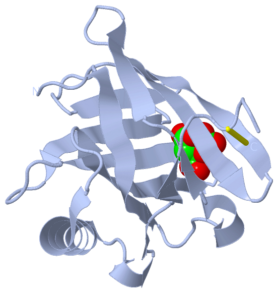
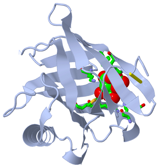
(-) Ligands, Modified Residues, Ions  (1, 1)

Asymmetric Unit (1, 1)
No.NameCountTypeFull Name
1FLC1Ligand/IonCITRATE ANION
Biological Unit 1 (1, 1)
No.NameCountTypeFull Name
1FLC1Ligand/IonCITRATE ANION
Biological Unit 2 (0, 0)
No.NameCountTypeFull Name
1FLC-1Ligand/IonCITRATE ANION
Biological Unit 3 (1, 1)
No.NameCountTypeFull Name
1FLC1Ligand/IonCITRATE ANION

(-) Sites  (1, 1)

Asymmetric Unit (1, 1)
No.NameEvidenceResiduesDescription
1AC1SOFTWAREARG A:29 , ARG A:55 , LYS A:62 , TYR A:78 , LYS A:82 , ARG A:103 , ARG A:108 , MET A:110 , HOH A:307 , ASP B:9BINDING SITE FOR RESIDUE FLC A 201

(-) SS Bonds  (2, 2)

Asymmetric Unit
No.Residues
1A:59 -A:151
2B:59 -B:151

(-) Cis Peptide Bonds  (1, 1)

Asymmetric Unit
No.Residues
1Phe B:25 -Pro B:26

 Sequence-Structure Mapping

(-) SAPs(SNPs)/Variants  (0, 0)

(no "SAP(SNP)/Variant" information available for 4RUN)

(-) PROSITE Motifs  (0, 0)

(no "PROSITE Motif" information available for 4RUN)

(-) Exons   (0, 0)

(no "Exon" information available for 4RUN)

(-) Sequences/Alignments

Asymmetric Unit
   Reformat: Number of residues per line =  ('0' or empty: single-line sequence representation)
  Number of residues per labelling interval =   
  UniProt sequence: complete  aligned part    
   Show mapping: SCOP domains CATH domains Pfam domains Secondary structure (by author)
SAPs(SNPs) PROSITE motifs Exons
(details for a mapped element are shown in a popup box when the mouse pointer rests over it)
Chain A from PDB  Type:PROTEIN  Length:147
                                                                                                                                                                                   
               SCOP domains --------------------------------------------------------------------------------------------------------------------------------------------------- SCOP domains
               CATH domains --------------------------------------------------------------------------------------------------------------------------------------------------- CATH domains
               Pfam domains --------------------------------------------------------------------------------------------------------------------------------------------------- Pfam domains
         Sec.struct. author ....eeeeeeeeee....hhhhh..ee..eeeee.....eeeeeeeee..eeeeeeeee.......eeee....eeeeeee......eeeeeeeee..eeeeeeeeee....eehhhhhhhhhhhhhh..hhh.ee..ee....... Sec.struct. author
                 SAPs(SNPs) --------------------------------------------------------------------------------------------------------------------------------------------------- SAPs(SNPs)
                    PROSITE --------------------------------------------------------------------------------------------------------------------------------------------------- PROSITE
                 Transcript --------------------------------------------------------------------------------------------------------------------------------------------------- Transcript
                 4run A   8 EDITGTWYVKAMVVDKDFPEDRRPRKVSPVKVTALGGGNLEATFTFMREDRCIQKKILMRKTEEPGKFSAYGGRKLIYLQELPGTDDYVFYSKDQRRGGLRYMGNLVGRNPNTNLEALEEFKKLVQHKGLSEEDIFMPLQTGSCVLE 154
                                    17        27        37        47        57        67        77        87        97       107       117       127       137       147       

Chain B from PDB  Type:PROTEIN  Length:152
                                                                                                                                                                                        
               SCOP domains -------------------------------------------------------------------------------------------------------------------------------------------------------- SCOP domains
               CATH domains -------------------------------------------------------------------------------------------------------------------------------------------------------- CATH domains
               Pfam domains -------------------------------------------------------------------------------------------------------------------------------------------------------- Pfam domains
         Sec.struct. author ....hhhhhheeeeeeeee............ee..eeeee.....eeeeeeeee..eeeeeeeee.......eeee....eeeeeee......eeeeeee......eeeeeeee.....hhhhhhhhhhhhhhh......eee.ee...... Sec.struct. author
                 SAPs(SNPs) -------------------------------------------------------------------------------------------------------------------------------------------------------- SAPs(SNPs)
                    PROSITE -------------------------------------------------------------------------------------------------------------------------------------------------------- PROSITE
                 Transcript -------------------------------------------------------------------------------------------------------------------------------------------------------- Transcript
                 4run B   2 SFTLEEEDITGTWYVKAMVVDKDFPEDRRPRKVSPVKVTALGGGNLEATFTFMREDRCIQKKILMRKTEEPGKFSAYGGRKLIYLQELPGTDDYVFYSKDQRRGGLRYMGNLVGRNPNTNLEALEEFKKLVQHKGLSEEDIFMPLQTGSCVL 153
                                    11        21        31        41        51        61        71        81        91       101       111       121       131       141       151  

   Legend:   → Mismatch (orange background)
  - → Gap (green background, '-', border residues have a numbering label)
    → Modified Residue (blue background, lower-case, 'x' indicates undefined single-letter code, labelled with number + name)
  x → Chemical Group (purple background, 'x', labelled with number + name, e.g. ACE or NH2)
  extra numbering lines below/above indicate numbering irregularities and modified residue names etc., number ends below/above '|'

 Classification and Annotation

(-) SCOP Domains  (0, 0)

(no "SCOP Domain" information available for 4RUN)

(-) CATH Domains  (0, 0)

(no "CATH Domain" information available for 4RUN)

(-) Pfam Domains  (0, 0)

(no "Pfam Domain" information available for 4RUN)

(-) Gene Ontology  (7, 7)

Asymmetric Unit(hide GO term definitions)

 Visualization

(-) Interactive Views

Asymmetric Unit
  Complete Structure
    Jena3D(integrated viewing of ligand, site, SAP, PROSITE, SCOP information)
    WebMol | AstexViewer[tm]@PDBe
(Java Applets, require no local installation except for Java; loading may be slow)
    STRAP
(Java WebStart application, automatic local installation, requires Java; full application with system access!)
    RasMol
(require local installation)
    Molscript (VRML)
(requires installation of a VRML viewer; select preferred view via VRML and generate a mono or stereo PDF format file)
 
  Ligands, Modified Residues, Ions
    FLC  [ RasMol | Jena3D ]  +environment [ RasMol | Jena3D ]
 
  Sites
    AC1  [ RasMol ]  +environment [ RasMol ]
 
  Cis Peptide Bonds
    Phe B:25 - Pro B:26   [ RasMol ]  
 
Biological Units
  Complete Structure
    Biological Unit 1  [ Jena3D ]
    Biological Unit 2  [ Jena3D ]
    Biological Unit 3  [ Jena3D ]

(-) Still Images

Jmol
  protein: cartoon or spacefill or dots and stick; nucleic acid: cartoon and stick; ligands: spacefill; active site: stick
Molscript
  protein, nucleic acid: cartoon; ligands: spacefill; active site: ball and stick

 Databases and Analysis Tools

(-) Databases

Access by PDB/NDB ID
  4run
    Family and Domain InformationProDom | SYSTERS
    General Structural InformationGlycoscienceDB | MMDB | NDB | OCA | PDB | PDBe | PDBj | PDBsum | PDBWiki | PQS | PROTEOPEDIA
    Orientation in MembranesOPM
    Protein SurfaceSURFACE
    Secondary StructureDSSP (structure derived) | HSSP (homology derived)
    Structural GenomicsGeneCensus
    Structural NeighboursCE | VAST
    Structure ClassificationCATH | Dali | SCOP
    Validation and Original DataBMRB Data View | BMRB Restraints Grid | EDS | PROCHECK | RECOORD | WHAT_CHECK
 
Access by UniProt ID/Accession number
  OBP2A_HUMAN | Q9NY56
    Comparative Protein Structure ModelsModBase
    Genomic InformationEnsembl
    Protein-protein InteractionDIP
    Sequence, Family and Domain InformationInterPro | Pfam | SMART | UniProtKB/SwissProt
 
Access by Enzyme Classificator   (EC Number)
  (no 'Enzyme Classificator' available)
    General Enzyme InformationBRENDA | EC-PDB | Enzyme | IntEnz
    PathwayKEGG | MetaCyc
 
Access by Disease Identifier   (MIM ID)
  (no 'MIM ID' available)
    Disease InformationOMIM
 
Access by GenAge ID
  (no 'GenAge ID' available)
    Age Related InformationGenAge

(-) Analysis Tools

Access by PDB/NDB ID
    Domain InformationXDom
    Interatomic Contacts of Structural UnitsCSU
    Ligand-protein ContactsLPC
    Protein CavitiescastP
    Sequence and Secondary StructurePDBCartoon
    Structure AlignmentSTRAP(Java WebStart application, automatic local installation, requires Java; full application with system access!)
    Structure and Sequence BrowserSTING
 
Access by UniProt ID/Accession number
  OBP2A_HUMAN | Q9NY56
    Protein Disorder PredictionDisEMBL | FoldIndex | GLOBPLOT (for more information see DisProt)

 Related Entries

(-) Entries Sharing at Least One Protein Chain (UniProt ID)

(no "Entries Sharing at Least One Protein Chain" available for 4RUN)

(-) Related Entries Specified in the PDB File

(no "Related Entries Specified in the PDB File" available for 4RUN)