Show PDB file:   
         Plain Text   HTML   (compressed file size)
QuickSearch:   
by PDB,NDB,UniProt,PROSITE Code or Search Term(s)  
(-)Asym./Biol. Unit
(-)Asym./Biol. Unit - sites
collapse expand < >
Image Asym./Biol. Unit
Asym./Biol. Unit  (Jmol Viewer)
Image Asym./Biol. Unit - sites
Asym./Biol. Unit - sites  (Jmol Viewer)

(-) Description

Title :  CRYSTAL STRUCTURE OF HUMAN GLUTAREDOXIN WITH MESNA
 
Authors :  J. Badger, V. Sridhar, C. Logan, F. H. Hausheer, V. L. Nienaber
Date :  04 Nov 14  (Deposition) - 01 Apr 15  (Release) - 01 Apr 15  (Revision)
Method :  X-RAY DIFFRACTION
Resolution :  1.08
Chains :  Asym./Biol. Unit :  A
Keywords :  Redox-Active Center, Electron Transport (Keyword Search: [Gene Ontology, PubMed, Web (Google))
 
Reference :  A. R. Parker, P. N. Petluru, V. L. Nienaber, J. Badger, B. D. Leverett, K. Jair, V. Sridhar, C. Logan, P. Y. Ayala, H. Kochat, F. H. Hausheer
Cysteine Specific Targeting Of The Functionally Distinct Peroxiredoxin And Glutaredoxin Proteins By The Investigational Disulfide Bnp7787.
Molecules V. 20 4928 2015
PubMed-ID: 25793542  |  Reference-DOI: 10.3390/MOLECULES20034928

(-) Compounds

Molecule 1 - GLUTAREDOXIN-1
    ChainsA
    EngineeredYES
    Expression SystemESCHERICHIA COLI
    Expression System StrainBL21
    Expression System Taxid511693
    GeneGLRX, GRX
    Organism CommonHUMAN
    Organism ScientificHOMO SAPIENS
    Organism Taxid9606
    SynonymTHIOLTRANSFERASE-1, TTASE-1

 Structural Features

(-) Chains, Units

  1
Asymmetric/Biological Unit A

Summary Information (see also Sequences/Alignments below)

(-) Ligands, Modified Residues, Ions  (1, 2)

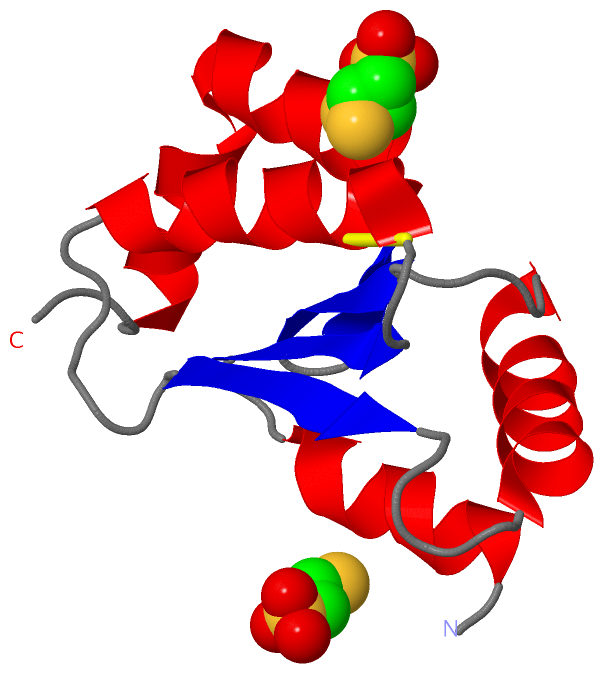
Asymmetric/Biological Unit (1, 2)
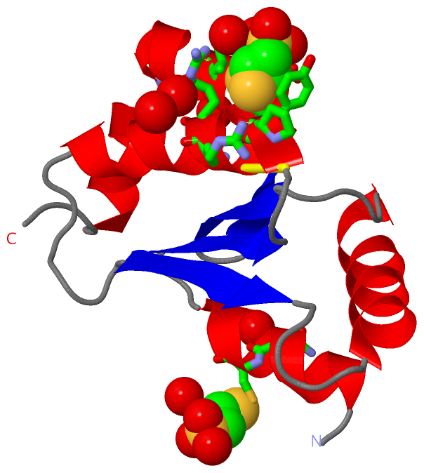
No.NameCountTypeFull Name
1COM2Ligand/Ion1-THIOETHANESULFONIC ACID

(-) Sites  (2, 2)

Asymmetric Unit (2, 2)
No.NameEvidenceResiduesDescription
1AC1SOFTWARECYS A:7 , PRO A:23 , ARG A:26 , ARG A:27 , GLU A:93 , HOH A:367 , HOH A:369 , HOH A:412 , HOH A:487BINDING SITE FOR RESIDUE COM A 201
2AC2SOFTWAREASN A:6 , TYR A:24 , ARG A:27 , CYS A:82 , SER A:83 , VAL A:86 , HOH A:328 , HOH A:347 , HOH A:383BINDING SITE FOR RESIDUE COM A 202

(-) SS Bonds  (1, 1)

Asymmetric/Biological Unit
No.Residues
1A:22 -A:25

(-) Cis Peptide Bonds  (1, 1)

Asymmetric/Biological Unit
No.Residues
1Val A:69 -Pro A:70

 Sequence-Structure Mapping

(-) SAPs(SNPs)/Variants  (0, 0)

(no "SAP(SNP)/Variant" information available for 4RQR)

(-) PROSITE Motifs  (0, 0)

(no "PROSITE Motif" information available for 4RQR)

(-) Exons   (0, 0)

(no "Exon" information available for 4RQR)

(-) Sequences/Alignments

Asymmetric/Biological Unit
   Reformat: Number of residues per line =  ('0' or empty: single-line sequence representation)
  Number of residues per labelling interval =   
  UniProt sequence: complete  aligned part    
   Show mapping: SCOP domains CATH domains Pfam domains Secondary structure (by author)
SAPs(SNPs) PROSITE motifs Exons
(details for a mapped element are shown in a popup box when the mouse pointer rests over it)
Chain A from PDB  Type:PROTEIN  Length:107
                                                                                                                                           
               SCOP domains ----------------------------------------------------------------------------------------------------------- SCOP domains
               CATH domains ----------------------------------------------------------------------------------------------------------- CATH domains
               Pfam domains ----------------------------------------------------------------------------------------------------------- Pfam domains
         Sec.struct. author .hhhhhhhhhh....eeeee...hhhhhhhhhhhhh.......eeeee.....hhhhhhhhhhhhhh.....eeee..eeeehhhhhhhhhhhhhhhhhhhhh.... Sec.struct. author
                 SAPs(SNPs) ----------------------------------------------------------------------------------------------------------- SAPs(SNPs)
                    PROSITE ----------------------------------------------------------------------------------------------------------- PROSITE
                 Transcript ----------------------------------------------------------------------------------------------------------- Transcript
                 4rqr A  -1 GTAQEFVNCKIQPGKVVVFIKPTCPYCRRAQEILSQLPIKQGLLEFVDITATNHTNEIQDYLQQLTGARTVPRVFIGKDCIGGCSDLVSLQQSGELLTRLKQIGALQ 105
                                     8        18        28        38        48        58        68        78        88        98       

   Legend:   → Mismatch (orange background)
  - → Gap (green background, '-', border residues have a numbering label)
    → Modified Residue (blue background, lower-case, 'x' indicates undefined single-letter code, labelled with number + name)
  x → Chemical Group (purple background, 'x', labelled with number + name, e.g. ACE or NH2)
  extra numbering lines below/above indicate numbering irregularities and modified residue names etc., number ends below/above '|'

 Classification and Annotation

(-) SCOP Domains  (0, 0)

(no "SCOP Domain" information available for 4RQR)

(-) CATH Domains  (0, 0)

(no "CATH Domain" information available for 4RQR)

(-) Pfam Domains  (0, 0)

(no "Pfam Domain" information available for 4RQR)

(-) Gene Ontology  (16, 16)

Asymmetric/Biological Unit(hide GO term definitions)

 Visualization

(-) Interactive Views

Asymmetric/Biological Unit
  Complete Structure
    Jena3D(integrated viewing of ligand, site, SAP, PROSITE, SCOP information)
    WebMol | AstexViewer[tm]@PDBe
(Java Applets, require no local installation except for Java; loading may be slow)
    STRAP
(Java WebStart application, automatic local installation, requires Java; full application with system access!)
    RasMol
(require local installation)
    Molscript (VRML)
(requires installation of a VRML viewer; select preferred view via VRML and generate a mono or stereo PDF format file)
 
  Ligands, Modified Residues, Ions
    COM  [ RasMol | Jena3D ]  +environment [ RasMol | Jena3D ]
 
  Sites
    AC1  [ RasMol ]  +environment [ RasMol ]
    AC2  [ RasMol ]  +environment [ RasMol ]
 
  Cis Peptide Bonds
    Val A:69 - Pro A:70   [ RasMol ]  
 

(-) Still Images

Jmol
  protein: cartoon or spacefill or dots and stick; nucleic acid: cartoon and stick; ligands: spacefill; active site: stick
Molscript
  protein, nucleic acid: cartoon; ligands: spacefill; active site: ball and stick

 Databases and Analysis Tools

(-) Databases

Access by PDB/NDB ID
  4rqr
    Family and Domain InformationProDom | SYSTERS
    General Structural InformationGlycoscienceDB | MMDB | NDB | OCA | PDB | PDBe | PDBj | PDBsum | PDBWiki | PQS | PROTEOPEDIA
    Orientation in MembranesOPM
    Protein SurfaceSURFACE
    Secondary StructureDSSP (structure derived) | HSSP (homology derived)
    Structural GenomicsGeneCensus
    Structural NeighboursCE | VAST
    Structure ClassificationCATH | Dali | SCOP
    Validation and Original DataBMRB Data View | BMRB Restraints Grid | EDS | PROCHECK | RECOORD | WHAT_CHECK
 
Access by UniProt ID/Accession number
  GLRX1_HUMAN | P35754
    Comparative Protein Structure ModelsModBase
    Genomic InformationEnsembl
    Protein-protein InteractionDIP
    Sequence, Family and Domain InformationInterPro | Pfam | SMART | UniProtKB/SwissProt
 
Access by Enzyme Classificator   (EC Number)
  (no 'Enzyme Classificator' available)
    General Enzyme InformationBRENDA | EC-PDB | Enzyme | IntEnz
    PathwayKEGG | MetaCyc
 
Access by Disease Identifier   (MIM ID)
  (no 'MIM ID' available)
    Disease InformationOMIM
 
Access by GenAge ID
  (no 'GenAge ID' available)
    Age Related InformationGenAge

(-) Analysis Tools

Access by PDB/NDB ID
    Domain InformationXDom
    Interatomic Contacts of Structural UnitsCSU
    Ligand-protein ContactsLPC
    Protein CavitiescastP
    Sequence and Secondary StructurePDBCartoon
    Structure AlignmentSTRAP(Java WebStart application, automatic local installation, requires Java; full application with system access!)
    Structure and Sequence BrowserSTING
 
Access by UniProt ID/Accession number
  GLRX1_HUMAN | P35754
    Protein Disorder PredictionDisEMBL | FoldIndex | GLOBPLOT (for more information see DisProt)

 Related Entries

(-) Entries Sharing at Least One Protein Chain (UniProt ID)

UniProtKB/Swiss-Prot
        GLRX1_HUMAN | P357541b4q 1jhb

(-) Related Entries Specified in the PDB File

(no "Related Entries Specified in the PDB File" available for 4RQR)