|
|
|
|
 Description
|
|
Compounds
|
||||||||||||||||||||||||||||||||||||||||||||||||||||||||||||||||||||||||||||||||||||||||||||||||||||||||||
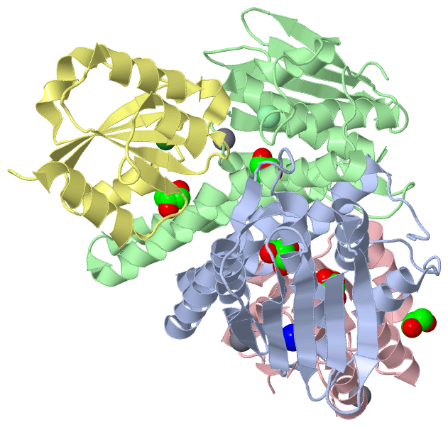
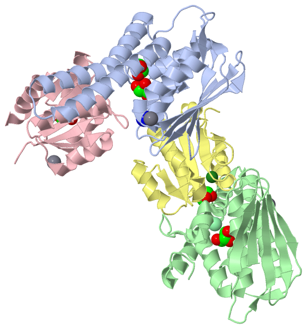
Chains, Units
Summary Information (see also Sequences/Alignments below) |
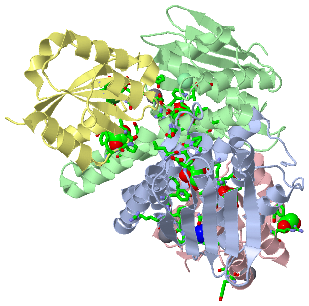
Ligands, Modified Residues, Ions (5, 11)
Asymmetric Unit (5, 11)
|
Sites (11, 11)
Asymmetric Unit (11, 11)
|
SS Bonds (0, 0)| (no "SS Bond" information available for 4QPJ) |
Cis Peptide Bonds (2, 2)
Asymmetric Unit
|
||||||||||||
SAPs(SNPs)/Variants (0, 0)| (no "SAP(SNP)/Variant" information available for 4QPJ) |
PROSITE Motifs (0, 0)| (no "PROSITE Motif" information available for 4QPJ) |
Exons (0, 0)| (no "Exon" information available for 4QPJ) |
Sequences/Alignments
Asymmetric Unit
Chain A from PDB Type:PROTEIN Length:208
SCOP domains ---------------------------------------------------------------------------------------------------------------------------------------------------------------------------------------------------------------- SCOP domains
CATH domains ---------------------------------------------------------------------------------------------------------------------------------------------------------------------------------------------------------------- CATH domains
Pfam domains ---------------------------------------------------------------------------------------------------------------------------------------------------------------------------------------------------------------- Pfam domains
SAPs(SNPs) ---------------------------------------------------------------------------------------------------------------------------------------------------------------------------------------------------------------- SAPs(SNPs)
PROSITE ---------------------------------------------------------------------------------------------------------------------------------------------------------------------------------------------------------------- PROSITE
Transcript ---------------------------------------------------------------------------------------------------------------------------------------------------------------------------------------------------------------- Transcript
4qpj A 2 SLPVTLSALDLGALLCSRICHDIISPIGAINNGLELLEEGGADEDAMALIKSSARNASARLQFARIAFGAAGSAGVQIDTGDAQNVATEYFRNEKPEFTWEGARVLLPKNKVKLLLNMLLIGNGAIPRGGSLAVRLEGSDTDPRFVITVKGRMLRVPPKFLELHSGAAPEEPIDAHSVQPYYTLLLAEEAGMKISIHATAEDIVFSAE 209
11 21 31 41 51 61 71 81 91 101 111 121 131 141 151 161 171 181 191 201
Chain B from PDB Type:PROTEIN Length:209
SCOP domains ----------------------------------------------------------------------------------------------------------------------------------------------------------------------------------------------------------------- SCOP domains
CATH domains ----------------------------------------------------------------------------------------------------------------------------------------------------------------------------------------------------------------- CATH domains
Pfam domains ----------------------------------------------------------------------------------------------------------------------------------------------------------------------------------------------------------------- Pfam domains
SAPs(SNPs) ----------------------------------------------------------------------------------------------------------------------------------------------------------------------------------------------------------------- SAPs(SNPs)
PROSITE ----------------------------------------------------------------------------------------------------------------------------------------------------------------------------------------------------------------- PROSITE
Transcript ----------------------------------------------------------------------------------------------------------------------------------------------------------------------------------------------------------------- Transcript
4qpj B 1 MSLPVTLSALDLGALLCSRICHDIISPIGAINNGLELLEEGGADEDAMALIKSSARNASARLQFARIAFGAAGSAGVQIDTGDAQNVATEYFRNEKPEFTWEGARVLLPKNKVKLLLNMLLIGNGAIPRGGSLAVRLEGSDTDPRFVITVKGRMLRVPPKFLELHSGAAPEEPIDAHSVQPYYTLLLAEEAGMKISIHATAEDIVFSAE 209
10 20 30 40 50 60 70 80 90 100 110 120 130 140 150 160 170 180 190 200
Chain C from PDB Type:PROTEIN Length:120
SCOP domains ------------------------------------------------------------------------------------------------------------------------ SCOP domains
CATH domains ------------------------------------------------------------------------------------------------------------------------ CATH domains
Pfam domains ------------------------------------------------------------------------------------------------------------------------ Pfam domains
SAPs(SNPs) ------------------------------------------------------------------------------------------------------------------------ SAPs(SNPs)
PROSITE ------------------------------------------------------------------------------------------------------------------------ PROSITE
Transcript ------------------------------------------------------------------------------------------------------------------------ Transcript
4qpj C -1 GSMRVLLIEDDSAIAQSIELMLKSESFNVYTTDLGEEGIDLGKLYDYDIILLDLNLPDMSGYEVLRTLRLSKVKTPILILSGMAGIEDKVRGLGFGADDYMTKPFHKDELIARIHAIVRR 118
8 18 28 38 48 58 68 78 88 98 108 118
Chain D from PDB Type:PROTEIN Length:121
SCOP domains ------------------------------------------------------------------------------------------------------------------------- SCOP domains
CATH domains ------------------------------------------------------------------------------------------------------------------------- CATH domains
Pfam domains ------------------------------------------------------------------------------------------------------------------------- Pfam domains
SAPs(SNPs) ------------------------------------------------------------------------------------------------------------------------- SAPs(SNPs)
PROSITE ------------------------------------------------------------------------------------------------------------------------- PROSITE
Transcript ------------------------------------------------------------------------------------------------------------------------- Transcript
4qpj D -2 RGSMRVLLIEDDSAIAQSIELMLKSESFNVYTTDLGEEGIDLGKLYDYDIILLDLNLPDMSGYEVLRTLRLSKVKTPILILSGMAGIEDKVRGLGFGADDYMTKPFHKDELIARIHAIVRR 118
7 17 27 37 47 57 67 77 87 97 107 117
|
||||||||||||||||||||
SCOP Domains (0, 0)| (no "SCOP Domain" information available for 4QPJ) |
CATH Domains (0, 0)| (no "CATH Domain" information available for 4QPJ) |
Pfam Domains (0, 0)| (no "Pfam Domain" information available for 4QPJ) |
Gene Ontology (5, 5)|
Asymmetric Unit(hide GO term definitions) |
Interactive Views
|
|||||||||||||||||||||||||||||||||||||||||||||||||||||||||||||||||||||||||||||||||||||||||||||||||||||||||||||||||||||||||||||||||||||||||||||||||||||||||||||||||||||||||||||||||||||||||||||||||||||||||||||||||||||||||||||||||||||||||||||||||||||||
Still Images
|
||||||||||||||||
Databases
|
||||||||||||||||||||||||||||||||||||||||||||||||||||||||||||||||||||||||||||||||||||||||||||||||||||||||||||||||||||||||||||||||||||||||||||||||||||||||||||||||||||||||||||||||||||||||||
Analysis Tools
|
||||||||||||||||||||||||||||||||||||||||||||||||||||||||||||||||||||||||
Entries Sharing at Least One Protein Chain (UniProt ID)
Related Entries Specified in the PDB File
|
|