|
|
|
|
 Description
|
|
Compounds
|
||||||||||||||||||||||||||||||||||||||||||||||||||||||||||||||||||||
Chains, Units
Summary Information (see also Sequences/Alignments below) |
Ligands, Modified Residues, Ions (1, 4)
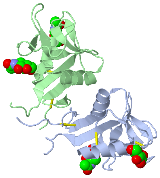
Asymmetric Unit (1, 4)
|
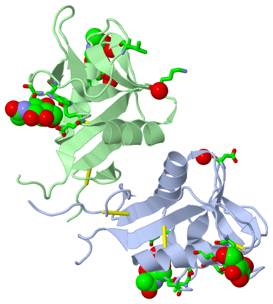
Sites (4, 4)
Asymmetric Unit (4, 4)
|
SS Bonds (6, 6)
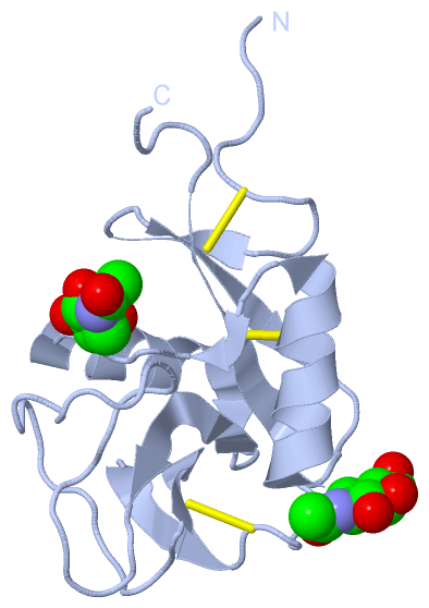
Asymmetric Unit
|
||||||||||||||||||||||||||||
Cis Peptide Bonds (0, 0)| (no "Cis Peptide Bond" information available for 4QKH) |
SAPs(SNPs)/Variants (0, 0)| (no "SAP(SNP)/Variant" information available for 4QKH) |
PROSITE Motifs (0, 0)| (no "PROSITE Motif" information available for 4QKH) |
Exons (0, 0)| (no "Exon" information available for 4QKH) |
Sequences/Alignments
Asymmetric Unit
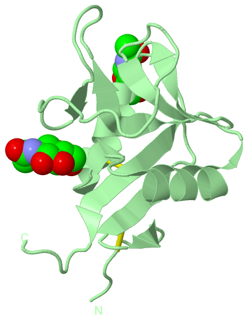
Chain A from PDB Type:PROTEIN Length:123
SCOP domains --------------------------------------------------------------------------------------------------------------------------- SCOP domains
CATH domains --------------------------------------------------------------------------------------------------------------------------- CATH domains
Pfam domains --------------------------------------------------------------------------------------------------------------------------- Pfam domains
SAPs(SNPs) --------------------------------------------------------------------------------------------------------------------------- SAPs(SNPs)
PROSITE --------------------------------------------------------------------------------------------------------------------------- PROSITE
Transcript --------------------------------------------------------------------------------------------------------------------------- Transcript
4qkh A 70 TGQAACPESWIGFQRKCFYFSDDTKNWTSSQRFCDSQDADLAQVESFQELNFLLRYKGPSDHWIGLSREQGQPWKWINGTEWTRQFPILGAGECAYLNDKGASSARCYTERKWICSKSDIHVG 192
79 89 99 109 119 129 139 149 159 169 179 189
Chain B from PDB Type:PROTEIN Length:121
SCOP domains ------------------------------------------------------------------------------------------------------------------------- SCOP domains
CATH domains ------------------------------------------------------------------------------------------------------------------------- CATH domains
Pfam domains ------------------------------------------------------------------------------------------------------------------------- Pfam domains
SAPs(SNPs) ------------------------------------------------------------------------------------------------------------------------- SAPs(SNPs)
PROSITE ------------------------------------------------------------------------------------------------------------------------- PROSITE
Transcript ------------------------------------------------------------------------------------------------------------------------- Transcript
4qkh B 72 QAACPESWIGFQRKCFYFSDDTKNWTSSQRFCDSQDADLAQVESFQELNFLLRYKGPSDHWIGLSREQGQPWKWINGTEWTRQFPILGAGECAYLNDKGASSARCYTERKWICSKSDIHVG 192
81 91 101 111 121 131 141 151 161 171 181 191
|
||||||||||||||||||||
SCOP Domains (0, 0)| (no "SCOP Domain" information available for 4QKH) |
CATH Domains (0, 0)| (no "CATH Domain" information available for 4QKH) |
Pfam Domains (0, 0)| (no "Pfam Domain" information available for 4QKH) |
Gene Ontology (10, 10)|
Asymmetric Unit(hide GO term definitions) |
Interactive Views
|
|||||||||||||||||||||||||||||||||||||||||||||||||||||||||||||||||||||||||||||||||||||||||||||||||||||||||||||||||||||||||||||||||||||||||||||||||||||||||||||||||||||||
Still Images
|
||||||||||||||||
Databases
|
||||||||||||||||||||||||||||||||||||||||||||||||||||||||||||||||||||||||||||||||||||||||||||||||||||||||||||||||||||||||||||||||||||||||||||||||||||||||||||||||
Analysis Tools
|
|||||||||||||||||||||||||||||||||||||||||||||||||||||||||||||
Entries Sharing at Least One Protein Chain (UniProt ID)
Related Entries Specified in the PDB File
|
|