Show PDB file:   
         Plain Text   HTML   (compressed file size)
QuickSearch:   
by PDB,NDB,UniProt,PROSITE Code or Search Term(s)  
(-)Asymmetric Unit
(-)Asym. Unit - sites
(-)Biological Unit 1
(-)Biol. Unit 1 - sites
(-)Biological Unit 2
collapse expand < >
Image Asymmetric Unit
Asymmetric Unit  (Jmol Viewer)
Image Asym. Unit - sites
Asym. Unit - sites  (Jmol Viewer)
Image Biological Unit 1
Biological Unit 1  (Jmol Viewer)
Image Biol. Unit 1 - sites
Biol. Unit 1 - sites  (Jmol Viewer)
Image Biological Unit 2
Biological Unit 2  (Jmol Viewer)

(-) Description

Title :  NEUTROPHIL SERINE PROTEASE 4 (PRSS57) APO FORM 1
 
Authors :  C. Eigenbrot, S. J. Lin, K. C. Dong
Date :  25 Apr 14  (Deposition) - 03 Sep 14  (Release) - 17 Sep 14  (Revision)
Method :  X-RAY DIFFRACTION
Resolution :  2.55
Chains :  Asym. Unit :  A,B
Biol. Unit 1:  A  (1x)
Biol. Unit 2:  B  (1x)
Keywords :  Trypsin Homology, Peptidase, Hydrolase (Keyword Search: [Gene Ontology, PubMed, Web (Google))
 
Reference :  S. J. Lin, K. C. Dong, C. Eigenbrot, M. Van Lookeren Campagne, D. Kirchhofer
Structures Of Neutrophil Serine Protease 4 Reveal An Unusua Mechanism Of Substrate Recognition By A Trypsin-Fold Protease.
Structure V. 22 1333 2014
PubMed-ID: 25156428  |  Reference-DOI: 10.1016/J.STR.2014.07.008

(-) Compounds

Molecule 1 - SERINE PROTEASE 57
    ChainsA, B
    EC Number3.4.21.-
    EngineeredYES
    Expression SystemSPODOPTERA FRUGIPERDA
    Expression System Taxid7108
    GenePRSS57, PRSSL1, UNQ782/PRO1599
    Organism CommonHUMAN
    Organism ScientificHOMO SAPIENS
    Organism Taxid9606
    SynonymSERINE PROTEASE 1-LIKE PROTEIN 1

 Structural Features

(-) Chains, Units

  12
Asymmetric Unit AB
Biological Unit 1 (1x)A 
Biological Unit 2 (1x) B

Summary Information (see also Sequences/Alignments below)

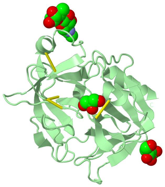
(-) Ligands, Modified Residues, Ions  (3, 9)

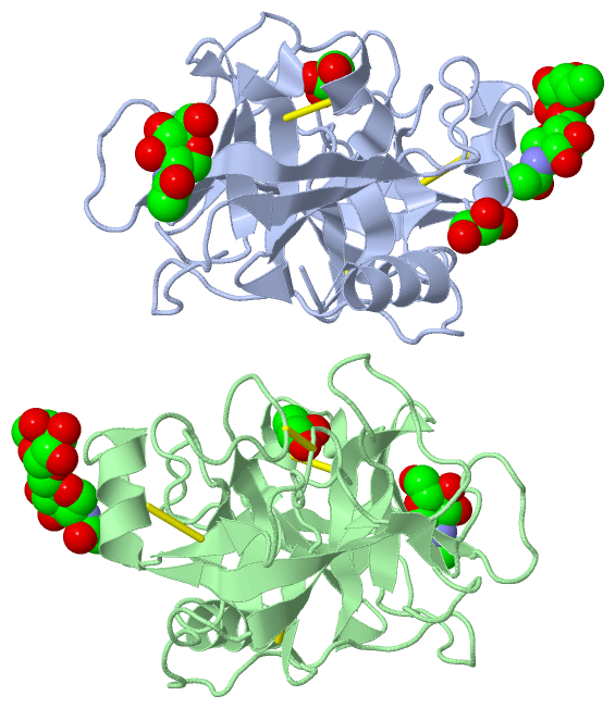
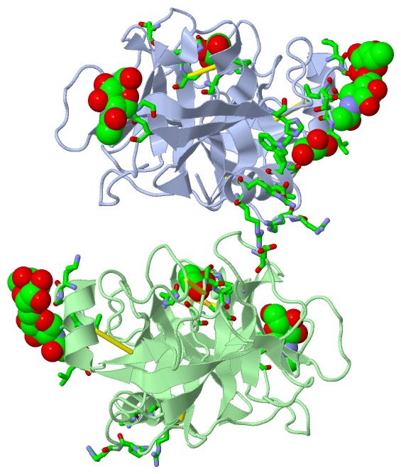
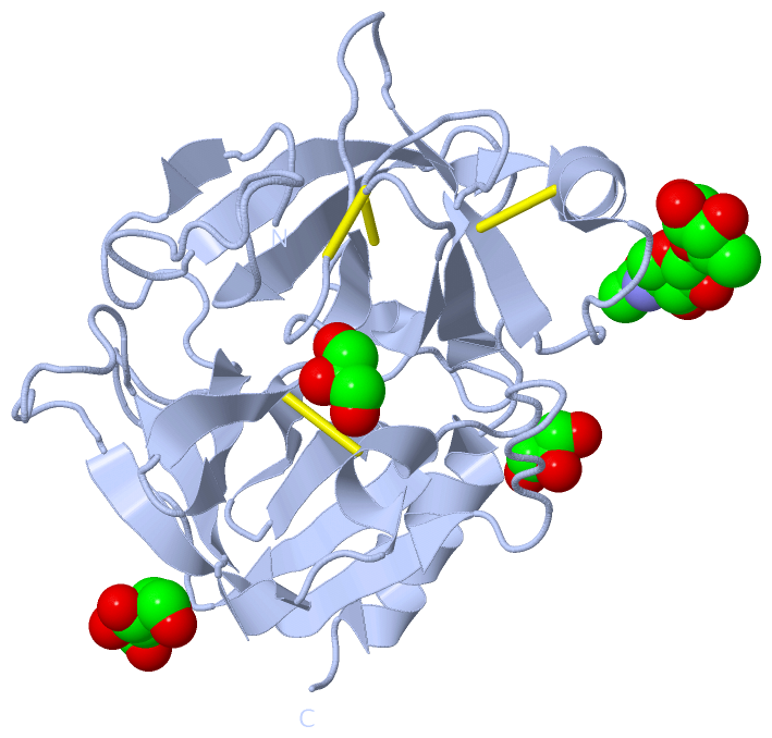
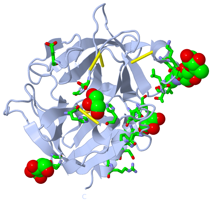
Asymmetric Unit (3, 9)
No.NameCountTypeFull Name
1FUC2Ligand/IonALPHA-L-FUCOSE
2GOL3Ligand/IonGLYCEROL
3NAG4Ligand/IonN-ACETYL-D-GLUCOSAMINE
Biological Unit 1 (3, 5)
No.NameCountTypeFull Name
1FUC1Ligand/IonALPHA-L-FUCOSE
2GOL2Ligand/IonGLYCEROL
3NAG2Ligand/IonN-ACETYL-D-GLUCOSAMINE
Biological Unit 2 (3, 4)
No.NameCountTypeFull Name
1FUC1Ligand/IonALPHA-L-FUCOSE
2GOL1Ligand/IonGLYCEROL
3NAG2Ligand/IonN-ACETYL-D-GLUCOSAMINE

(-) Sites  (9, 9)

Asymmetric Unit (9, 9)
No.NameEvidenceResiduesDescription
1AC1SOFTWAREGLU A:150 , ASN A:169 , GLY A:174 , LEU A:176 , THR A:177 , LEU A:178 , FUC A:302 , ARG B:125 , ARG B:126 , ALA B:236 , ASP B:240 , ARG B:243BINDING SITE FOR RESIDUE NAG A 301
2AC2SOFTWARELYS A:173 , NAG A:301 , ARG B:243 , ARG B:244BINDING SITE FOR RESIDUE FUC A 302
3AC3SOFTWAREGLY A:84 , ASN A:109BINDING SITE FOR RESIDUE NAG A 303
4AC4SOFTWAREHIS A:41 , CYS A:42 , HIS A:57 , SER A:192 , ALA A:193 , SER A:195BINDING SITE FOR RESIDUE GOL A 304
5AC5SOFTWAREHIS A:91 , ALA A:233 , PHE A:234 , TRP A:237BINDING SITE FOR RESIDUE GOL A 305
6AC6SOFTWAREARG A:125 , ARG A:126 , ALA A:236 , ASP A:240 , ARG A:243 , GLU B:150 , ASN B:169 , GLY B:174 , LEU B:176 , THR B:177 , LEU B:178 , FUC B:302BINDING SITE FOR RESIDUE NAG B 301
7AC7SOFTWAREARG A:243 , ARG A:244 , LYS B:173 , NAG B:301BINDING SITE FOR RESIDUE FUC B 302
8AC8SOFTWAREGLY B:84 , ASN B:109BINDING SITE FOR RESIDUE NAG B 303
9AC9SOFTWAREHIS B:41 , CYS B:42 , HIS B:57 , SER B:192 , ALA B:193 , SER B:195BINDING SITE FOR RESIDUE GOL B 304

(-) SS Bonds  (8, 8)

Asymmetric Unit
No.Residues
1A:42 -A:58
2A:136 -A:201
3A:168 -A:182
4A:191 -A:220
5B:42 -B:58
6B:136 -B:201
7B:168 -B:182
8B:191 -B:220

(-) Cis Peptide Bonds  (0, 0)

(no "Cis Peptide Bond" information available for 4Q7X)

 Sequence-Structure Mapping

(-) SAPs(SNPs)/Variants  (0, 0)

(no "SAP(SNP)/Variant" information available for 4Q7X)

(-) PROSITE Motifs  (0, 0)

(no "PROSITE Motif" information available for 4Q7X)

(-) Exons   (0, 0)

(no "Exon" information available for 4Q7X)

(-) Sequences/Alignments

Asymmetric Unit
   Reformat: Number of residues per line =  ('0' or empty: single-line sequence representation)
  Number of residues per labelling interval =   
  UniProt sequence: complete  aligned part    
   Show mapping: SCOP domains CATH domains Pfam domains Secondary structure (by author)
SAPs(SNPs) PROSITE motifs Exons
(details for a mapped element are shown in a popup box when the mouse pointer rests over it)
Chain A from PDB  Type:PROTEIN  Length:235
                                                                                                                                                                                                                                                                            
               SCOP domains ------------------------------------------------------------------------------------------------------------------------------------------------------------------------------------------------------------------------------------------- SCOP domains
               CATH domains ------------------------------------------------------------------------------------------------------------------------------------------------------------------------------------------------------------------------------------------- CATH domains
               Pfam domains ------------------------------------------------------------------------------------------------------------------------------------------------------------------------------------------------------------------------------------------- Pfam domains
         Sec.struct. author ....ee........eeeeee..eeeeeeeeee..eeeehhhhhh..hhh.eeeee............eeeeeeeeee.............eeeee...........................eeeeee..............eeeeeeeehhhhhhhhh.......eeeee................eeee..eeeeeeee...........eeeee...hhhhhhhhhhhh... Sec.struct. author
                 SAPs(SNPs) ------------------------------------------------------------------------------------------------------------------------------------------------------------------------------------------------------------------------------------------- SAPs(SNPs)
                    PROSITE ------------------------------------------------------------------------------------------------------------------------------------------------------------------------------------------------------------------------------------------- PROSITE
                 Transcript ------------------------------------------------------------------------------------------------------------------------------------------------------------------------------------------------------------------------------------------- Transcript
                4q7x A   16 IIGGHEVTPHSRPYMASVRFGGQHHCGGFLLRARWVVSAAHCFSHRDLRTGLVVLGAHVLSTAEPTQQVFGIDALTTHPDYHPMTHANDICLLRLNGSAVLGPAVGLLRLPGRRARPPTAGTRCRVAGWGFVSDFEELPPGLMEAKVRVLDPDVCNSSWKGHLTLTMLCTRSGDSHRRGFCSADSGGPLVCRNRAHGLVSFSGLWCGDPKTPDVYTQVSAFVAWIWDVVRRSSPQ  248
                                    25        35||      46        56      ||63        73        83        93       103       113       123 |     132       142  ||   153       163       173       183   ||  190       200 ||    214       223       233       243     
                                               36|                      62A||                                                           124A                  145|                                    186A|  |           202|            222A                          
                                                38                       62B|                                                                                  147                                     186B  |            207                                          
                                                                          62C                                                                                                                             188A                                                         

Chain B from PDB  Type:PROTEIN  Length:232
                                                                                                                                                                                                                                                                         
               SCOP domains ---------------------------------------------------------------------------------------------------------------------------------------------------------------------------------------------------------------------------------------- SCOP domains
               CATH domains ---------------------------------------------------------------------------------------------------------------------------------------------------------------------------------------------------------------------------------------- CATH domains
               Pfam domains ---------------------------------------------------------------------------------------------------------------------------------------------------------------------------------------------------------------------------------------- Pfam domains
         Sec.struct. author ....ee........eeeeee..eeeeeeeeee..eeeehhhhhh..hhh.eeeee............eee.eeeeee.............eeeee...........................eeeeee..............eeeeeeeehhhhhhhhh.......eeeee................eeee..eeeeeeee...........eeeee...hhhhhhhhhhhh Sec.struct. author
                 SAPs(SNPs) ---------------------------------------------------------------------------------------------------------------------------------------------------------------------------------------------------------------------------------------- SAPs(SNPs)
                    PROSITE ---------------------------------------------------------------------------------------------------------------------------------------------------------------------------------------------------------------------------------------- PROSITE
                 Transcript ---------------------------------------------------------------------------------------------------------------------------------------------------------------------------------------------------------------------------------------- Transcript
                4q7x B   16 IIGGHEVTPHSRPYMASVRFGGQHHCGGFLLRARWVVSAAHCFSHRDLRTGLVVLGAHVLSTAEPTQQVFGIDALTTHPDYHPMTHANDICLLRLNGSAVLGPAVGLLRLPGRRARPPTAGTRCRVAGWGFVSDFEELPPGLMEAKVRVLDPDVCNSSWKGHLTLTMLCTRSGDSHRRGFCSADSGGPLVCRNRAHGLVSFSGLWCGDPKTPDVYTQVSAFVAWIWDVVRRS  245
                                    25        35||      46        56      ||63        73        83        93       103       113       123 |     132       142  ||   153       163       173       183   ||  190       200 ||    214       223       233       243  
                                               36|                      62A||                                                           124A                  145|                                    186A|  |           202|            222A                       
                                                38                       62B|                                                                                  147                                     186B  |            207                                       
                                                                          62C                                                                                                                             188A                                                      

   Legend:   → Mismatch (orange background)
  - → Gap (green background, '-', border residues have a numbering label)
    → Modified Residue (blue background, lower-case, 'x' indicates undefined single-letter code, labelled with number + name)
  x → Chemical Group (purple background, 'x', labelled with number + name, e.g. ACE or NH2)
  extra numbering lines below/above indicate numbering irregularities and modified residue names etc., number ends below/above '|'

 Classification and Annotation

(-) SCOP Domains  (0, 0)

(no "SCOP Domain" information available for 4Q7X)

(-) CATH Domains  (0, 0)

(no "CATH Domain" information available for 4Q7X)

(-) Pfam Domains  (0, 0)

(no "Pfam Domain" information available for 4Q7X)

(-) Gene Ontology  (6, 6)

Asymmetric Unit(hide GO term definitions)

 Visualization

(-) Interactive Views

Asymmetric Unit
  Complete Structure
    Jena3D(integrated viewing of ligand, site, SAP, PROSITE, SCOP information)
    WebMol | AstexViewer[tm]@PDBe
(Java Applets, require no local installation except for Java; loading may be slow)
    STRAP
(Java WebStart application, automatic local installation, requires Java; full application with system access!)
    RasMol
(require local installation)
    Molscript (VRML)
(requires installation of a VRML viewer; select preferred view via VRML and generate a mono or stereo PDF format file)
 
  Ligands, Modified Residues, Ions
    FUC  [ RasMol | Jena3D ]  +environment [ RasMol | Jena3D ]
    GOL  [ RasMol | Jena3D ]  +environment [ RasMol | Jena3D ]
    NAG  [ RasMol | Jena3D ]  +environment [ RasMol | Jena3D ]
 
  Sites
    AC1  [ RasMol ]  +environment [ RasMol ]
    AC2  [ RasMol ]  +environment [ RasMol ]
    AC3  [ RasMol ]  +environment [ RasMol ]
    AC4  [ RasMol ]  +environment [ RasMol ]
    AC5  [ RasMol ]  +environment [ RasMol ]
    AC6  [ RasMol ]  +environment [ RasMol ]
    AC7  [ RasMol ]  +environment [ RasMol ]
    AC8  [ RasMol ]  +environment [ RasMol ]
    AC9  [ RasMol ]  +environment [ RasMol ]
 
  Cis Peptide Bonds
(no "Cis Peptide Bonds" information available for 4q7x)
 
Biological Units
  Complete Structure
    Biological Unit 1  [ Jena3D ]
    Biological Unit 2  [ Jena3D ]

(-) Still Images

Jmol
  protein: cartoon or spacefill or dots and stick; nucleic acid: cartoon and stick; ligands: spacefill; active site: stick
Molscript
  protein, nucleic acid: cartoon; ligands: spacefill; active site: ball and stick

 Databases and Analysis Tools

(-) Databases

Access by PDB/NDB ID
  4q7x
    Family and Domain InformationProDom | SYSTERS
    General Structural InformationGlycoscienceDB | MMDB | NDB | OCA | PDB | PDBe | PDBj | PDBsum | PDBWiki | PQS | PROTEOPEDIA
    Orientation in MembranesOPM
    Protein SurfaceSURFACE
    Secondary StructureDSSP (structure derived) | HSSP (homology derived)
    Structural GenomicsGeneCensus
    Structural NeighboursCE | VAST
    Structure ClassificationCATH | Dali | SCOP
    Validation and Original DataBMRB Data View | BMRB Restraints Grid | EDS | PROCHECK | RECOORD | WHAT_CHECK
 
Access by UniProt ID/Accession number
  PRS57_HUMAN | Q6UWY2
    Comparative Protein Structure ModelsModBase
    Genomic InformationEnsembl
    Protein-protein InteractionDIP
    Sequence, Family and Domain InformationInterPro | Pfam | SMART | UniProtKB/SwissProt
 
Access by Enzyme Classificator   (EC Number)
  3.4.21.-
    General Enzyme InformationBRENDA | EC-PDB | Enzyme | IntEnz
    PathwayKEGG | MetaCyc
 
Access by Disease Identifier   (MIM ID)
  (no 'MIM ID' available)
    Disease InformationOMIM
 
Access by GenAge ID
  (no 'GenAge ID' available)
    Age Related InformationGenAge

(-) Analysis Tools

Access by PDB/NDB ID
    Domain InformationXDom
    Interatomic Contacts of Structural UnitsCSU
    Ligand-protein ContactsLPC
    Protein CavitiescastP
    Sequence and Secondary StructurePDBCartoon
    Structure AlignmentSTRAP(Java WebStart application, automatic local installation, requires Java; full application with system access!)
    Structure and Sequence BrowserSTING
 
Access by UniProt ID/Accession number
  PRS57_HUMAN | Q6UWY2
    Protein Disorder PredictionDisEMBL | FoldIndex | GLOBPLOT (for more information see DisProt)

 Related Entries

(-) Entries Sharing at Least One Protein Chain (UniProt ID)

UniProtKB/Swiss-Prot
        PRS57_HUMAN | Q6UWY24q7y 4q7z 4q80

(-) Related Entries Specified in the PDB File

4q7y 4q7z 4q80