Show PDB file:   
         Plain Text   HTML   (compressed file size)
QuickSearch:   
by PDB,NDB,UniProt,PROSITE Code or Search Term(s)  
(-)Asymmetric Unit
(-)Asym. Unit - sites
(-)Biological Unit 1
(-)Biol. Unit 1 - sites
(-)Biological Unit 2
collapse expand < >
Image Asymmetric Unit
Asymmetric Unit  (Jmol Viewer)
Image Asym. Unit - sites
Asym. Unit - sites  (Jmol Viewer)
Image Biological Unit 1
Biological Unit 1  (Jmol Viewer)
Image Biol. Unit 1 - sites
Biol. Unit 1 - sites  (Jmol Viewer)
Image Biological Unit 2
Biological Unit 2  (Jmol Viewer)

(-) Description

Title :  BROMODOMAIN OF TRYPANOSOMA BRUCEI BDF2 WITH IBET-151
 
Authors :  E. W. Debler
Date :  15 May 14  (Deposition) - 18 Nov 15  (Release) - 18 Nov 15  (Revision)
Method :  X-RAY DIFFRACTION
Resolution :  1.25
Chains :  Asym. Unit :  A,B
Biol. Unit 1:  A  (1x)
Biol. Unit 2:  B  (1x)
Keywords :  Signaling Protein, Inhibitor, Histone, Epigenetic Reader, Signaling Protein-Inhibitor Complex (Keyword Search: [Gene Ontology, PubMed, Web (Google))
 
Reference :  D. Schulz, M. Mugnier, E. Paulsen, H. Kim, C. Chung, D. F. Tough, I. Rioja, R. K. Prinjha, N. Papavasiliou, E. W. Debler
Bromodomain Proteins Regulate Mono- Allelic Expression And Switching Of T. Brucei Variant Surface Glycoprotein
To Be Published
PubMed: search

(-) Compounds

Molecule 1 - BROMODOMAIN FACTOR 2
    ChainsA, B
    EngineeredYES
    Expression SystemESCHERICHIA COLI
    Expression System Taxid562
    FragmentBROMODOMAIN, UNP RESIDUES 9-123
    GeneTB10.6K15.3240
    Organism ScientificTRYPANOSOMA BRUCEI BRUCEI
    Organism Taxid999953
    Strain927/4 GUTAT10.1

 Structural Features

(-) Chains, Units

  12
Asymmetric Unit AB
Biological Unit 1 (1x)A 
Biological Unit 2 (1x) B

Summary Information (see also Sequences/Alignments below)

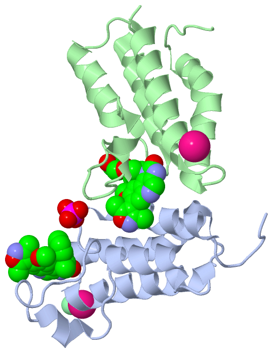
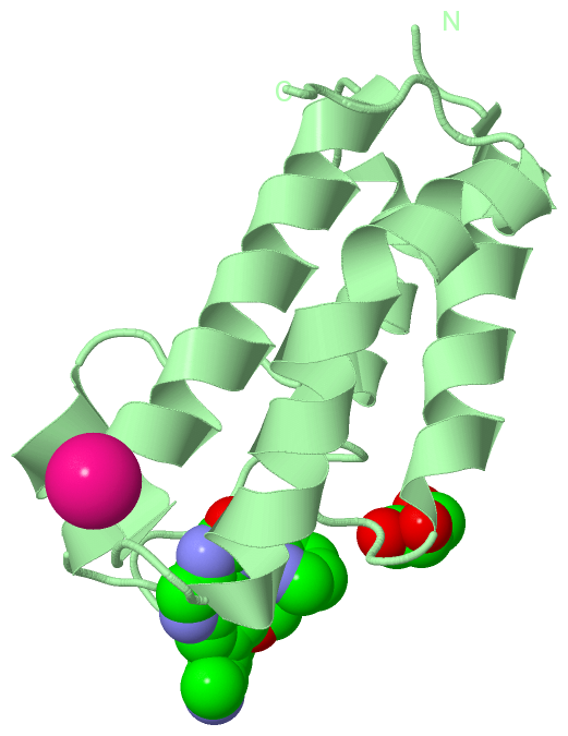
(-) Ligands, Modified Residues, Ions  (5, 7)

Asymmetric Unit (5, 7)
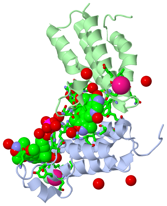
No.NameCountTypeFull Name
11GH2Ligand/Ion7-(3,5-DIMETHYL-1,2-OXAZOL-4-YL)-8-METHOXY-1-[(1R)-1-(PYRIDIN-2-YL)ETHYL]-1H,2H,3H-IMIDAZO[4,5-C]QUINOLIN-2-ONE
2CL1Ligand/IonCHLORIDE ION
3GOL1Ligand/IonGLYCEROL
4K2Ligand/IonPOTASSIUM ION
5PO41Ligand/IonPHOSPHATE ION
Biological Unit 1 (2, 2)
No.NameCountTypeFull Name
11GH1Ligand/Ion7-(3,5-DIMETHYL-1,2-OXAZOL-4-YL)-8-METHOXY-1-[(1R)-1-(PYRIDIN-2-YL)ETHYL]-1H,2H,3H-IMIDAZO[4,5-C]QUINOLIN-2-ONE
2CL-1Ligand/IonCHLORIDE ION
3GOL-1Ligand/IonGLYCEROL
4K-1Ligand/IonPOTASSIUM ION
5PO41Ligand/IonPHOSPHATE ION
Biological Unit 2 (2, 2)
No.NameCountTypeFull Name
11GH1Ligand/Ion7-(3,5-DIMETHYL-1,2-OXAZOL-4-YL)-8-METHOXY-1-[(1R)-1-(PYRIDIN-2-YL)ETHYL]-1H,2H,3H-IMIDAZO[4,5-C]QUINOLIN-2-ONE
2CL-1Ligand/IonCHLORIDE ION
3GOL1Ligand/IonGLYCEROL
4K-1Ligand/IonPOTASSIUM ION
5PO4-1Ligand/IonPHOSPHATE ION

(-) Sites  (7, 7)

Asymmetric Unit (7, 7)
No.NameEvidenceResiduesDescription
1AC1SOFTWARELEU A:28 , GLY A:29 , MET A:30 , VAL A:35 , GLU A:39 , TYR A:85 , ASN A:86 , TRP A:92 , ASP A:105 , GLN A:109 , HOH A:321 , HOH A:348 , HOH A:388 , GLN B:94binding site for residue 1GH A 201
2AC2SOFTWARETRP A:23 , HIS A:32 , HIS A:33 , HOH A:352 , HOH A:391 , HOH A:432 , HOH A:435 , HOH B:397binding site for residue PO4 A 202
3AC3SOFTWARELEU A:83 , GLU A:84 , ASN A:86 , HOH A:360 , HOH A:420 , HOH B:334binding site for residue K A 203
4AC4SOFTWARELYS A:88 , HOH A:380binding site for residue CL A 204
5AC5SOFTWARELEU A:16 , VAL A:19 , SER A:20 , TRP A:23 , ARG A:57 , HOH A:302 , LEU B:28 , GLY B:29 , MET B:30 , GLU B:39 , LEU B:40 , ASN B:86 , TRP B:92 , HOH B:365 , HOH B:373binding site for residue 1GH B 201
6AC6SOFTWAREHOH A:341 , LEU B:83 , GLU B:84 , ASN B:86 , HOH B:348 , HOH B:391binding site for residue K B 202
7AC7SOFTWAREASP A:24 , LYS A:27 , GLY B:29 , HIS B:32 , HIS B:33 , HOH B:318 , HOH B:390binding site for residue GOL B 203

(-) SS Bonds  (0, 0)

(no "SS Bond" information available for 4PKL)

(-) Cis Peptide Bonds  (0, 0)

(no "Cis Peptide Bond" information available for 4PKL)

 Sequence-Structure Mapping

(-) SAPs(SNPs)/Variants  (0, 0)

(no "SAP(SNP)/Variant" information available for 4PKL)

(-) PROSITE Motifs  (0, 0)

(no "PROSITE Motif" information available for 4PKL)

(-) Exons   (0, 0)

(no "Exon" information available for 4PKL)

(-) Sequences/Alignments

Asymmetric Unit
   Reformat: Number of residues per line =  ('0' or empty: single-line sequence representation)
  Number of residues per labelling interval =   
  UniProt sequence: complete  aligned part    
   Show mapping: SCOP domains CATH domains Pfam domains Secondary structure (by author)
SAPs(SNPs) PROSITE motifs Exons
(details for a mapped element are shown in a popup box when the mouse pointer rests over it)
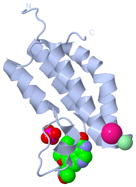
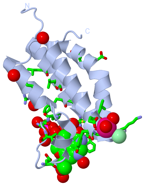
Chain A from PDB  Type:PROTEIN  Length:110
                                                                                                                                              
               SCOP domains -------------------------------------------------------------------------------------------------------------- SCOP domains
               CATH domains -------------------------------------------------------------------------------------------------------------- CATH domains
               Pfam domains -------------------------------------------------------------------------------------------------------------- Pfam domains
         Sec.struct. author ......hhhhhhhhhhhhhhhh...............hhhhhh....hhhhhhhhhhhh...hhhhhhhhhhhhhhhhhhhh...hhhhhhhhhhhhhhhhhhhhhh... Sec.struct. author
                 SAPs(SNPs) -------------------------------------------------------------------------------------------------------------- SAPs(SNPs)
                    PROSITE -------------------------------------------------------------------------------------------------------------- PROSITE
                 Transcript -------------------------------------------------------------------------------------------------------------- Transcript
                 4pkl A   5 GPHMSFNKNGCLVFVSRLWDLDKLGMFHHPVSAEELPDYHTVIKRPVDLSSIRDGIEKGTYATDVDVQNDVARMITNALEYNAKGSTWYQEAMSFRKTYLDLARQSGLVV 114
                                    14        24        34        44        54        64        74        84        94       104       114

Chain B from PDB  Type:PROTEIN  Length:108
                                                                                                                                            
               SCOP domains ------------------------------------------------------------------------------------------------------------ SCOP domains
               CATH domains ------------------------------------------------------------------------------------------------------------ CATH domains
               Pfam domains ------------------------------------------------------------------------------------------------------------ Pfam domains
         Sec.struct. author ..hhhhhhhhhhhhhhhh...............hhhhhh....hhhhhhhhhhhh...hhhhhhhhhhhhhhhhhhhh...hhhhhhhhhhhhhhhhhhhhh...... Sec.struct. author
                 SAPs(SNPs) ------------------------------------------------------------------------------------------------------------ SAPs(SNPs)
                    PROSITE ------------------------------------------------------------------------------------------------------------ PROSITE
                 Transcript ------------------------------------------------------------------------------------------------------------ Transcript
                 4pkl B   9 SFNKNGCLVFVSRLWDLDKLGMFHHPVSAEELPDYHTVIKRPVDLSSIRDGIEKGTYATDVDVQNDVARMITNALEYNAKGSTWYQEAMSFRKTYLDLARQSGLVVDD 116
                                    18        28        38        48        58        68        78        88        98       108        

   Legend:   → Mismatch (orange background)
  - → Gap (green background, '-', border residues have a numbering label)
    → Modified Residue (blue background, lower-case, 'x' indicates undefined single-letter code, labelled with number + name)
  x → Chemical Group (purple background, 'x', labelled with number + name, e.g. ACE or NH2)
  extra numbering lines below/above indicate numbering irregularities and modified residue names etc., number ends below/above '|'

 Classification and Annotation

(-) SCOP Domains  (0, 0)

(no "SCOP Domain" information available for 4PKL)

(-) CATH Domains  (0, 0)

(no "CATH Domain" information available for 4PKL)

(-) Pfam Domains  (0, 0)

(no "Pfam Domain" information available for 4PKL)

(-) Gene Ontology  (4, 4)

Asymmetric Unit(hide GO term definitions)

 Visualization

(-) Interactive Views

Asymmetric Unit
  Complete Structure
    Jena3D(integrated viewing of ligand, site, SAP, PROSITE, SCOP information)
    WebMol | AstexViewer[tm]@PDBe
(Java Applets, require no local installation except for Java; loading may be slow)
    STRAP
(Java WebStart application, automatic local installation, requires Java; full application with system access!)
    RasMol
(require local installation)
    Molscript (VRML)
(requires installation of a VRML viewer; select preferred view via VRML and generate a mono or stereo PDF format file)
 
  Ligands, Modified Residues, Ions
    1GH  [ RasMol | Jena3D ]  +environment [ RasMol | Jena3D ]
    CL  [ RasMol | Jena3D ]  +environment [ RasMol | Jena3D ]
    GOL  [ RasMol | Jena3D ]  +environment [ RasMol | Jena3D ]
    K  [ RasMol | Jena3D ]  +environment [ RasMol | Jena3D ]
    PO4  [ RasMol | Jena3D ]  +environment [ RasMol | Jena3D ]
 
  Sites
    AC1  [ RasMol ]  +environment [ RasMol ]
    AC2  [ RasMol ]  +environment [ RasMol ]
    AC3  [ RasMol ]  +environment [ RasMol ]
    AC4  [ RasMol ]  +environment [ RasMol ]
    AC5  [ RasMol ]  +environment [ RasMol ]
    AC6  [ RasMol ]  +environment [ RasMol ]
    AC7  [ RasMol ]  +environment [ RasMol ]
 
  Cis Peptide Bonds
(no "Cis Peptide Bonds" information available for 4pkl)
 
Biological Units
  Complete Structure
    Biological Unit 1  [ Jena3D ]
    Biological Unit 2  [ Jena3D ]

(-) Still Images

Jmol
  protein: cartoon or spacefill or dots and stick; nucleic acid: cartoon and stick; ligands: spacefill; active site: stick
Molscript
  protein, nucleic acid: cartoon; ligands: spacefill; active site: ball and stick

 Databases and Analysis Tools

(-) Databases

Access by PDB/NDB ID
  4pkl
    Family and Domain InformationProDom | SYSTERS
    General Structural InformationGlycoscienceDB | MMDB | NDB | OCA | PDB | PDBe | PDBj | PDBsum | PDBWiki | PQS | PROTEOPEDIA
    Orientation in MembranesOPM
    Protein SurfaceSURFACE
    Secondary StructureDSSP (structure derived) | HSSP (homology derived)
    Structural GenomicsGeneCensus
    Structural NeighboursCE | VAST
    Structure ClassificationCATH | Dali | SCOP
    Validation and Original DataBMRB Data View | BMRB Restraints Grid | EDS | PROCHECK | RECOORD | WHAT_CHECK
 
Access by UniProt ID/Accession number
  Q38AM1_TRYB2 | Q38AM1
    Comparative Protein Structure ModelsModBase
    Genomic InformationEnsembl
    Protein-protein InteractionDIP
    Sequence, Family and Domain InformationInterPro | Pfam | SMART | UniProtKB/TrEMBL
 
Access by Enzyme Classificator   (EC Number)
  (no 'Enzyme Classificator' available)
    General Enzyme InformationBRENDA | EC-PDB | Enzyme | IntEnz
    PathwayKEGG | MetaCyc
 
Access by Disease Identifier   (MIM ID)
  (no 'MIM ID' available)
    Disease InformationOMIM
 
Access by GenAge ID
  (no 'GenAge ID' available)
    Age Related InformationGenAge

(-) Analysis Tools

Access by PDB/NDB ID
    Domain InformationXDom
    Interatomic Contacts of Structural UnitsCSU
    Ligand-protein ContactsLPC
    Protein CavitiescastP
    Sequence and Secondary StructurePDBCartoon
    Structure AlignmentSTRAP(Java WebStart application, automatic local installation, requires Java; full application with system access!)
    Structure and Sequence BrowserSTING
 
Access by UniProt ID/Accession number
  Q38AM1_TRYB2 | Q38AM1
    Protein Disorder PredictionDisEMBL | FoldIndex | GLOBPLOT (for more information see DisProt)

 Related Entries

(-) Entries Sharing at Least One Protein Chain (UniProt ID)

UniProtKB/TrEMBL
        Q38AM1_TRYB2 | Q38AM12n9g

(-) Related Entries Specified in the PDB File

(no "Related Entries Specified in the PDB File" available for 4PKL)