Show PDB file:   
         Plain Text   HTML   (compressed file size)
QuickSearch:   
by PDB,NDB,UniProt,PROSITE Code or Search Term(s)  
(-)Asym./Biol. Unit
(-)Asym./Biol. Unit - sites
collapse expand < >
Image Asym./Biol. Unit
Asym./Biol. Unit  (Jmol Viewer)
Image Asym./Biol. Unit - sites
Asym./Biol. Unit - sites  (Jmol Viewer)

(-) Description

Title :  STRUCTURE OF ESCHERICHIA COLI PGAB C-TERMINAL DOMAIN, P212121 CRYSTAL FORM
 
Authors :  D. J. Little, G. Li, C. Ing, B. Difrancesco, N. C. Bamford, H. Robinson, M R. Pomes, P. L. Howell
Date :  27 Mar 14  (Deposition) - 02 Jul 14  (Release) - 01 Oct 14  (Revision)
Method :  X-RAY DIFFRACTION
Resolution :  1.80
Chains :  Asym./Biol. Unit :  A
Keywords :  Beta Alpha Barrel, Carbohydrate Binding, Glycosyl Hydrolase Fold (Keyword Search: [Gene Ontology, PubMed, Web (Google))
 
Reference :  D. J. Little, G. Li, C. Ing, B. R. Difrancesco, N. C. Bamford, H. Robinson, M. Nitz, R. Pomes, P. L. Howell
Modification And Periplasmic Translocation Of The Biofilm Exopolysaccharide Poly-Beta-1, 6-N-Acetyl-D-Glucosamine.
Proc. Natl. Acad. Sci. Usa V. 111 11013 2014
PubMed-ID: 24994902  |  Reference-DOI: 10.1073/PNAS.1406388111

(-) Compounds

Molecule 1 - POLY-BETA-1,6-N-ACETYL-D-GLUCOSAMINE N-DEACETYLASE
    ChainsA
    EC Number3.5.1.-
    EngineeredYES
    Expression SystemESCHERICHIA COLI BL21
    Expression System PlasmidPET28A
    Expression System StrainBL21
    Expression System Taxid511693
    Expression System Vector TypePLASMID
    FragmentC-TERMINAL DOMAIN (UNP RESIDUES 310-672)
    GenePGAB, YCDR, B1023, JW5142
    Organism ScientificESCHERICHIA COLI K12
    Organism Taxid83333
    StrainK12
    SynonymPGAB, POLY-BETA-1,6-GLCNAC N-DEACETYLASE

 Structural Features

(-) Chains, Units

  1
Asymmetric/Biological Unit A

Summary Information (see also Sequences/Alignments below)

(-) Ligands, Modified Residues, Ions  (2, 2)

Asymmetric/Biological Unit (2, 2)
No.NameCountTypeFull Name
1CL1Ligand/IonCHLORIDE ION
2EDO1Ligand/Ion1,2-ETHANEDIOL

(-) Sites  (2, 2)

Asymmetric Unit (2, 2)
No.NameEvidenceResiduesDescription
1AC1SOFTWAREARG A:342 , TRP A:387 , PHE A:649 , ASN A:652 , HOH A:987binding site for residue EDO A 701
2AC2SOFTWAREALA A:478binding site for residue CL A 702

(-) SS Bonds  (0, 0)

(no "SS Bond" information available for 4P7L)

(-) Cis Peptide Bonds  (4, 4)

Asymmetric/Biological Unit
No.Residues
1Ser A:312 -Pro A:313
2Glu A:474 -Asp A:475
3Tyr A:645 -Pro A:646
4Gln A:653 -Pro A:654

 Sequence-Structure Mapping

(-) SAPs(SNPs)/Variants  (0, 0)

(no "SAP(SNP)/Variant" information available for 4P7L)

(-) PROSITE Motifs  (0, 0)

(no "PROSITE Motif" information available for 4P7L)

(-) Exons   (0, 0)

(no "Exon" information available for 4P7L)

(-) Sequences/Alignments

Asymmetric/Biological Unit
   Reformat: Number of residues per line =  ('0' or empty: single-line sequence representation)
  Number of residues per labelling interval =   
  UniProt sequence: complete  aligned part    
   Show mapping: SCOP domains CATH domains Pfam domains Secondary structure (by author)
SAPs(SNPs) PROSITE motifs Exons
(details for a mapped element are shown in a popup box when the mouse pointer rests over it)
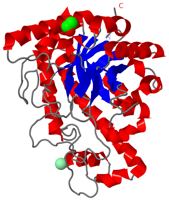
Chain A from PDB  Type:PROTEIN  Length:357
                                                                                                                                                                                                                                                                                                                                                                                                     
               SCOP domains --------------------------------------------------------------------------------------------------------------------------------------------------------------------------------------------------------------------------------------------------------------------------------------------------------------------------------------------------------------------- SCOP domains
               CATH domains --------------------------------------------------------------------------------------------------------------------------------------------------------------------------------------------------------------------------------------------------------------------------------------------------------------------------------------------------------------------- CATH domains
               Pfam domains --------------------------------------------------------------------------------------------------------------------------------------------------------------------------------------------------------------------------------------------------------------------------------------------------------------------------------------------------------------------- Pfam domains
         Sec.struct. author .....eeeee........hhhhhhhhhhhhhhhhhhhh..eeeee..........................hhhhhhhhhhhh...eeeeee.....................................hhhhhhhhhhhhhhhhhhh...eeee............hhhhhhhhhhh....hhhhhh.hhhhhhhhhhhhhhhhhhhhhhhhhhhhhhhh...eeeeeehhhhhhhhhhhhhh..hhhhhhhhh.eeeee........hhhhhhhhhhhhhhhhh....hhh.eeeeee.eee.....eee.hhhhhhhhhhhhhhh....eeee..hhhhh..hhhhhhhhhh.. Sec.struct. author
                 SAPs(SNPs) --------------------------------------------------------------------------------------------------------------------------------------------------------------------------------------------------------------------------------------------------------------------------------------------------------------------------------------------------------------------- SAPs(SNPs)
                    PROSITE --------------------------------------------------------------------------------------------------------------------------------------------------------------------------------------------------------------------------------------------------------------------------------------------------------------------------------------------------------------------- PROSITE
                 Transcript --------------------------------------------------------------------------------------------------------------------------------------------------------------------------------------------------------------------------------------------------------------------------------------------------------------------------------------------------------------------- Transcript
                 4p7l A 310 EKSPQRIMHIDLDYVYDENLQQMDRNIDVLIQRVKDMQISTVYLQAFADPDGDGLVKEVWFPNRLLPMKADIFSRVAWQLRTRSGVNIYAWMPVLSWDLDPTLTRVKYLPTGEKKAQIHPEQYHRLSPFDDRVRAQVGMLYEDLAGHAAFDGILFHDDALLSDYEDASAPAITAYQQAGFSGSLSEIRQNPEQFKQWARFKSRALTDFTLELSARVKAIRGPHIKTARNIFALPVIQPESEAWFAQNYADFLKSYDWTAIMAMPYLEGVAEKSADQWLIQLTNQIKNIPQAKDKSILELQAQNWQKNGQHQAISSQQLAHWMSLLQLNGVKNYGYYPDNFLHNQPEIDLIRPEFSTA 666
                                   319       329       339       349       359       369       379       389       399       409       419       429       439       449       459       469       479       489       499       509       519       529       539       549       559       569       579       589       599       609       619       629       639       649       659       

   Legend:   → Mismatch (orange background)
  - → Gap (green background, '-', border residues have a numbering label)
    → Modified Residue (blue background, lower-case, 'x' indicates undefined single-letter code, labelled with number + name)
  x → Chemical Group (purple background, 'x', labelled with number + name, e.g. ACE or NH2)
  extra numbering lines below/above indicate numbering irregularities and modified residue names etc., number ends below/above '|'

 Classification and Annotation

(-) SCOP Domains  (0, 0)

(no "SCOP Domain" information available for 4P7L)

(-) CATH Domains  (0, 0)

(no "CATH Domain" information available for 4P7L)

(-) Pfam Domains  (0, 0)

(no "Pfam Domain" information available for 4P7L)

(-) Gene Ontology  (9, 9)

Asymmetric/Biological Unit(hide GO term definitions)

 Visualization

(-) Interactive Views

Asymmetric/Biological Unit
  Complete Structure
    Jena3D(integrated viewing of ligand, site, SAP, PROSITE, SCOP information)
    WebMol | AstexViewer[tm]@PDBe
(Java Applets, require no local installation except for Java; loading may be slow)
    STRAP
(Java WebStart application, automatic local installation, requires Java; full application with system access!)
    RasMol
(require local installation)
    Molscript (VRML)
(requires installation of a VRML viewer; select preferred view via VRML and generate a mono or stereo PDF format file)
 
  Ligands, Modified Residues, Ions
    CL  [ RasMol | Jena3D ]  +environment [ RasMol | Jena3D ]
    EDO  [ RasMol | Jena3D ]  +environment [ RasMol | Jena3D ]
 
  Sites
    AC1  [ RasMol ]  +environment [ RasMol ]
    AC2  [ RasMol ]  +environment [ RasMol ]
 
  Cis Peptide Bonds
    Gln A:653 - Pro A:654   [ RasMol ]  
    Glu A:474 - Asp A:475   [ RasMol ]  
    Ser A:312 - Pro A:313   [ RasMol ]  
    Tyr A:645 - Pro A:646   [ RasMol ]  
 

(-) Still Images

Jmol
  protein: cartoon or spacefill or dots and stick; nucleic acid: cartoon and stick; ligands: spacefill; active site: stick
Molscript
  protein, nucleic acid: cartoon; ligands: spacefill; active site: ball and stick

 Databases and Analysis Tools

(-) Databases

Access by PDB/NDB ID
  4p7l
    Family and Domain InformationProDom | SYSTERS
    General Structural InformationGlycoscienceDB | MMDB | NDB | OCA | PDB | PDBe | PDBj | PDBsum | PDBWiki | PQS | PROTEOPEDIA
    Orientation in MembranesOPM
    Protein SurfaceSURFACE
    Secondary StructureDSSP (structure derived) | HSSP (homology derived)
    Structural GenomicsGeneCensus
    Structural NeighboursCE | VAST
    Structure ClassificationCATH | Dali | SCOP
    Validation and Original DataBMRB Data View | BMRB Restraints Grid | EDS | PROCHECK | RECOORD | WHAT_CHECK
 
Access by UniProt ID/Accession number
  PGAB_ECOLI | P75906
    Comparative Protein Structure ModelsModBase
    Genomic InformationEnsembl
    Protein-protein InteractionDIP
    Sequence, Family and Domain InformationInterPro | Pfam | SMART | UniProtKB/SwissProt
 
Access by Enzyme Classificator   (EC Number)
  3.5.1.-
    General Enzyme InformationBRENDA | EC-PDB | Enzyme | IntEnz
    PathwayKEGG | MetaCyc
 
Access by Disease Identifier   (MIM ID)
  (no 'MIM ID' available)
    Disease InformationOMIM
 
Access by GenAge ID
  (no 'GenAge ID' available)
    Age Related InformationGenAge

(-) Analysis Tools

Access by PDB/NDB ID
    Domain InformationXDom
    Interatomic Contacts of Structural UnitsCSU
    Ligand-protein ContactsLPC
    Protein CavitiescastP
    Sequence and Secondary StructurePDBCartoon
    Structure AlignmentSTRAP(Java WebStart application, automatic local installation, requires Java; full application with system access!)
    Structure and Sequence BrowserSTING
 
Access by UniProt ID/Accession number
  PGAB_ECOLI | P75906
    Protein Disorder PredictionDisEMBL | FoldIndex | GLOBPLOT (for more information see DisProt)

 Related Entries

(-) Entries Sharing at Least One Protein Chain (UniProt ID)

UniProtKB/Swiss-Prot
        PGAB_ECOLI | P759063vus 4f9d 4f9j 4p7n 4p7o 4p7q 4p7r

(-) Related Entries Specified in the PDB File

4f9d PGAB RESIDUES 42-655 IN COMPLEX WITH NICKEL
4f9j PGAB RESIDUES 42-655 IN COMPLEX WITH IRON
4p7n
4p7o
4p7q
4p7r