Show PDB file:   
         Plain Text   HTML   (compressed file size)
QuickSearch:   
by PDB,NDB,UniProt,PROSITE Code or Search Term(s)  
(-)Asymmetric Unit
(-)Asym. Unit - sites
(-)Biological Unit 1
(-)Biol. Unit 1 - sites
collapse expand < >
Image Asymmetric Unit
Asymmetric Unit  (Jmol Viewer)
Image Asym. Unit - sites
Asym. Unit - sites  (Jmol Viewer)
Image Biological Unit 1
Biological Unit 1  (Jmol Viewer)
Image Biol. Unit 1 - sites
Biol. Unit 1 - sites  (Jmol Viewer)

(-) Description

Title :  DIRECTED EVOLUTION OF A B3 METALLO-BETA-LACTAMASE AIM-1
 
Authors :  C. -F. D. Hou, C. Collyer, D. L. Ollis
Date :  21 Mar 14  (Deposition) - 27 May 15  (Release) - 27 May 15  (Revision)
Method :  X-RAY DIFFRACTION
Resolution :  1.89
Chains :  Asym. Unit :  A
Biol. Unit 1:  A  (2x)
Keywords :  Metallo-Beta-Lactamase, Antibiotic Resistance, Directed Evolution, Hydrolase (Keyword Search: [Gene Ontology, PubMed, Web (Google))
 
Reference :  C. -F. D. Hou, C. Collyer, D. L. Ollis
Directed Evolution Of A B3 Metallo-Beta-Lactamase Aim-1
To Be Published
PubMed: search

(-) Compounds

Molecule 1 - METALLO-BETA-LACTAMASE AIM-1
    ChainsA
    EngineeredYES
    Expression SystemESCHERICHIA COLI
    Expression System Taxid562
    GeneBLAAIM-1
    Organism ScientificPSEUDOMONAS AERUGINOSA
    Organism Taxid287

 Structural Features

(-) Chains, Units

  1
Asymmetric Unit A
Biological Unit 1 (2x)A

Summary Information (see also Sequences/Alignments below)
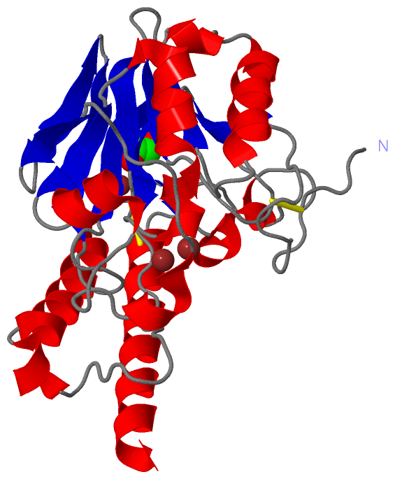
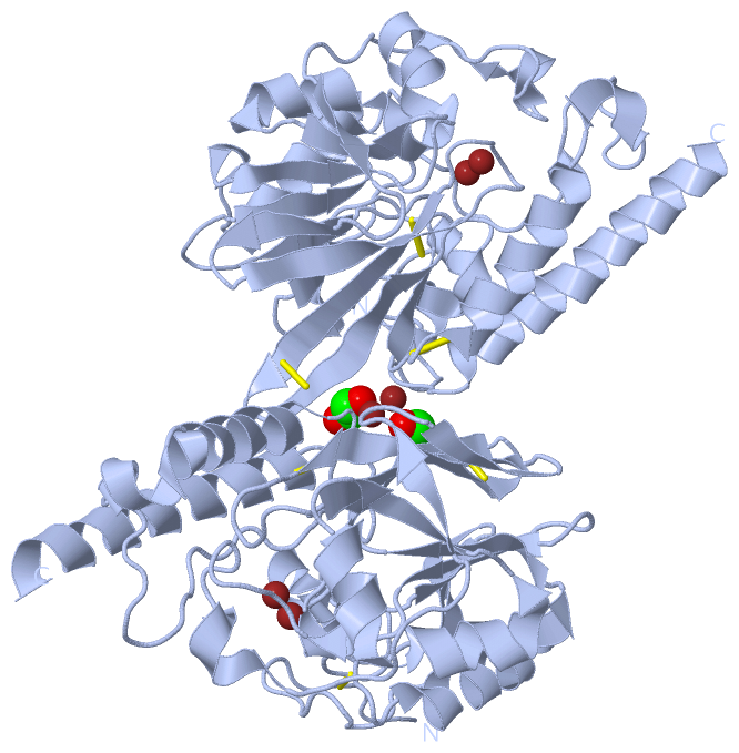
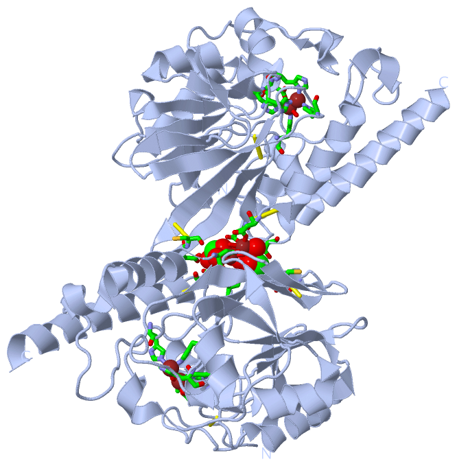
(-) Ligands, Modified Residues, Ions  (2, 4)

Asymmetric Unit (2, 4)
No.NameCountTypeFull Name
1EDO1Ligand/Ion1,2-ETHANEDIOL
2ZN3Ligand/IonZINC ION
Biological Unit 1 (1, 2)
No.NameCountTypeFull Name
1EDO2Ligand/Ion1,2-ETHANEDIOL
2ZN-1Ligand/IonZINC ION

(-) Sites  (4, 4)

Asymmetric Unit (4, 4)
No.NameEvidenceResiduesDescription
1AC1SOFTWAREHIS A:104 , HIS A:106 , HIS A:182 , ZN A:302 , HOH A:581 , HOH A:605binding site for residue ZN A 301
2AC2SOFTWAREASP A:108 , HIS A:109 , HIS A:250 , ZN A:301 , HOH A:581 , HOH A:605binding site for residue ZN A 302
3AC3SOFTWAREASP A:242 , ASP A:244 , EDO A:304 , HOH A:402 , HOH A:408 , HOH A:424binding site for residue ZN A 303
4AC4SOFTWAREASP A:198 , CYS A:199 , ASP A:244 , THR A:271 , ZN A:303 , HOH A:402 , HOH A:408 , HOH A:424binding site for residue EDO A 304

(-) SS Bonds  (3, 3)

Asymmetric Unit
No.Residues
1A:31 -A:56
2A:194 -A:199
3A:243 -A:274

(-) Cis Peptide Bonds  (0, 0)

(no "Cis Peptide Bond" information available for 4P62)

 Sequence-Structure Mapping

(-) SAPs(SNPs)/Variants  (0, 0)

(no "SAP(SNP)/Variant" information available for 4P62)

(-) PROSITE Motifs  (0, 0)

(no "PROSITE Motif" information available for 4P62)

(-) Exons   (0, 0)

(no "Exon" information available for 4P62)

(-) Sequences/Alignments

Asymmetric Unit
   Reformat: Number of residues per line =  ('0' or empty: single-line sequence representation)
  Number of residues per labelling interval =   
  UniProt sequence: complete  aligned part    
   Show mapping: SCOP domains CATH domains Pfam domains Secondary structure (by author)
SAPs(SNPs) PROSITE motifs Exons
(details for a mapped element are shown in a popup box when the mouse pointer rests over it)
Chain A from PDB  Type:PROTEIN  Length:269
                                                                                                                                                                                                                                                                                                             
               SCOP domains ----------------------------------------------------------------------------------------------------------------------------------------------------------------------------------------------------------------------------------------------------------------------------- SCOP domains
               CATH domains ----------------------------------------------------------------------------------------------------------------------------------------------------------------------------------------------------------------------------------------------------------------------------- CATH domains
               Pfam domains ----------------------------------------------------------------------------------------------------------------------------------------------------------------------------------------------------------------------------------------------------------------------------- Pfam domains
         Sec.struct. author .................eeee..eee........eee.....eeee......hhhhhhhhhhhh..hhh.eeeee....hhhhhhhhhhhhhhhh..eeee..hhhhhhhh.......hhhhh........eeee....eeee..eeeeeee........eeeeeeeee..eeeeeee...............hhhhh.hhhhhhhhhhhhhhh....eee..hhhhhhhhhh............hhhhhhhhhhhhhhhhhhhhhhhh Sec.struct. author
                 SAPs(SNPs) ----------------------------------------------------------------------------------------------------------------------------------------------------------------------------------------------------------------------------------------------------------------------------- SAPs(SNPs)
                    PROSITE ----------------------------------------------------------------------------------------------------------------------------------------------------------------------------------------------------------------------------------------------------------------------------- PROSITE
                 Transcript ----------------------------------------------------------------------------------------------------------------------------------------------------------------------------------------------------------------------------------------------------------------------------- Transcript
                 4p62 A  27 ASRGCADDAGWNDPAMPQKVYGNTWYVGTCGISALLVTSDAGHILVDAATPQAGPQILANIRALGFRPEDVRAIVLSHEHFDHAGSLAELQKATGAPVYARAPAIDTLKRGLPDRTDPQFEVAEPVAPVANIVTLADDGVVSVGPLALTAVASPGHTPGGTSWTWRSCEGDDCRQMVYADSLTAISDDVYRYSDDAAHPGYLAAFRNTLARVAALDCDILVTPHPSASGLWNRIGPRAAAPLMDTTACRRYAQGARQRLEKRLAEEAAA 295
                                    36        46        56        66        76        86        96       106       116       126       136       146       156       166       176       186       196       206       216       226       236       246       256       266       276       286         

   Legend:   → Mismatch (orange background)
  - → Gap (green background, '-', border residues have a numbering label)
    → Modified Residue (blue background, lower-case, 'x' indicates undefined single-letter code, labelled with number + name)
  x → Chemical Group (purple background, 'x', labelled with number + name, e.g. ACE or NH2)
  extra numbering lines below/above indicate numbering irregularities and modified residue names etc., number ends below/above '|'

 Classification and Annotation

(-) SCOP Domains  (0, 0)

(no "SCOP Domain" information available for 4P62)

(-) CATH Domains  (0, 0)

(no "CATH Domain" information available for 4P62)

(-) Pfam Domains  (0, 0)

(no "Pfam Domain" information available for 4P62)

(-) Gene Ontology  (1, 1)

Asymmetric Unit(hide GO term definitions)

 Visualization

(-) Interactive Views

Asymmetric Unit
  Complete Structure
    Jena3D(integrated viewing of ligand, site, SAP, PROSITE, SCOP information)
    WebMol | AstexViewer[tm]@PDBe
(Java Applets, require no local installation except for Java; loading may be slow)
    STRAP
(Java WebStart application, automatic local installation, requires Java; full application with system access!)
    RasMol
(require local installation)
    Molscript (VRML)
(requires installation of a VRML viewer; select preferred view via VRML and generate a mono or stereo PDF format file)
 
  Ligands, Modified Residues, Ions
    EDO  [ RasMol | Jena3D ]  +environment [ RasMol | Jena3D ]
    ZN  [ RasMol | Jena3D ]  +environment [ RasMol | Jena3D ]
 
  Sites
    AC1  [ RasMol ]  +environment [ RasMol ]
    AC2  [ RasMol ]  +environment [ RasMol ]
    AC3  [ RasMol ]  +environment [ RasMol ]
    AC4  [ RasMol ]  +environment [ RasMol ]
 
  Cis Peptide Bonds
(no "Cis Peptide Bonds" information available for 4p62)
 
Biological Unit
  Complete Structure
    Biological Unit 1  [ Jena3D ]

(-) Still Images

Jmol
  protein: cartoon or spacefill or dots and stick; nucleic acid: cartoon and stick; ligands: spacefill; active site: stick
Molscript
  protein, nucleic acid: cartoon; ligands: spacefill; active site: ball and stick

 Databases and Analysis Tools

(-) Databases

Access by PDB/NDB ID
  4p62
    Family and Domain InformationProDom | SYSTERS
    General Structural InformationGlycoscienceDB | MMDB | NDB | OCA | PDB | PDBe | PDBj | PDBsum | PDBWiki | PQS | PROTEOPEDIA
    Orientation in MembranesOPM
    Protein SurfaceSURFACE
    Secondary StructureDSSP (structure derived) | HSSP (homology derived)
    Structural GenomicsGeneCensus
    Structural NeighboursCE | VAST
    Structure ClassificationCATH | Dali | SCOP
    Validation and Original DataBMRB Data View | BMRB Restraints Grid | EDS | PROCHECK | RECOORD | WHAT_CHECK
 
Access by UniProt ID/Accession number
  B5DCA0_PSEAI | B5DCA0
    Comparative Protein Structure ModelsModBase
    Genomic InformationEnsembl
    Protein-protein InteractionDIP
    Sequence, Family and Domain InformationInterPro | Pfam | SMART | UniProtKB/TrEMBL
 
Access by Enzyme Classificator   (EC Number)
  (no 'Enzyme Classificator' available)
    General Enzyme InformationBRENDA | EC-PDB | Enzyme | IntEnz
    PathwayKEGG | MetaCyc
 
Access by Disease Identifier   (MIM ID)
  (no 'MIM ID' available)
    Disease InformationOMIM
 
Access by GenAge ID
  (no 'GenAge ID' available)
    Age Related InformationGenAge

(-) Analysis Tools

Access by PDB/NDB ID
    Domain InformationXDom
    Interatomic Contacts of Structural UnitsCSU
    Ligand-protein ContactsLPC
    Protein CavitiescastP
    Sequence and Secondary StructurePDBCartoon
    Structure AlignmentSTRAP(Java WebStart application, automatic local installation, requires Java; full application with system access!)
    Structure and Sequence BrowserSTING
 
Access by UniProt ID/Accession number
  B5DCA0_PSEAI | B5DCA0
    Protein Disorder PredictionDisEMBL | FoldIndex | GLOBPLOT (for more information see DisProt)

 Related Entries

(-) Entries Sharing at Least One Protein Chain (UniProt ID)

UniProtKB/TrEMBL
        B5DCA0_PSEAI | B5DCA04awy 4awz 4ax0 4ax1

(-) Related Entries Specified in the PDB File

(no "Related Entries Specified in the PDB File" available for 4P62)