Show PDB file:   
         Plain Text   HTML   (compressed file size)
QuickSearch:   
by PDB,NDB,UniProt,PROSITE Code or Search Term(s)  
(-)Asymmetric Unit
(-)Asym. Unit - sites
(-)Biological Unit 1
(-)Biol. Unit 1 - sites
(-)Biological Unit 2
collapse expand < >
Image Asymmetric Unit
Asymmetric Unit  (Jmol Viewer)
Image Asym. Unit - sites
Asym. Unit - sites  (Jmol Viewer)
Image Biological Unit 1
Biological Unit 1  (Jmol Viewer)
Image Biol. Unit 1 - sites
Biol. Unit 1 - sites  (Jmol Viewer)
Image Biological Unit 2
Biological Unit 2  (Jmol Viewer)

(-) Description

Title :  CRYSTAL STRUCTURE OF RAT DNPH1 (RCL) WITH 6-NAPHTHYL-PURINE-RIBOSIDE-MONOPHOSPHATE
 
Authors :  A. Padilla, G. Labesse, P. A. Kaminski
Date :  16 Mar 14  (Deposition) - 20 Aug 14  (Release) - 20 Aug 14  (Revision)
Method :  X-RAY DIFFRACTION
Resolution :  2.11
Chains :  Asym. Unit :  A,C
Biol. Unit 1:  C  (2x)
Biol. Unit 2:  A  (2x)
Keywords :  N-Glycosidase, Inhibitor, Hydrolase (Keyword Search: [Gene Ontology, PubMed, Web (Google))
 
Reference :  C. Amiable, J. Paoletti, A. Haouz, A. Padilla, G. Labesse, P. A. Kaminski, S. Pochet
6-(Hetero)Arylpurine Nucleotides As Inhibitors Of The Oncogenic Target Dnph1: Synthesis, Structural Studies And Cytotoxic Activities.
Eur. J. Med. Chem. V. 85C 418 2014
PubMed-ID: 25108359  |  Reference-DOI: 10.1016/J.EJMECH.2014.07.110
(for further references see the PDB file header)

(-) Compounds

Molecule 1 - 2'-DEOXYNUCLEOSIDE 5'-PHOSPHATE N-HYDROLASE 1
    ChainsC, A
    EC Number3.2.2.-
    EngineeredYES
    Expression SystemESCHERICHIA COLI
    Expression System PlasmidPET24A
    Expression System StrainBLI5
    Expression System Taxid562
    Expression System Vector TypePLASMID
    FragmentRESIDUES 11-151
    GeneDNPH1, RCL
    MutationYES
    Organism CommonRAT
    Organism ScientificRATTUS NORVEGICUS
    Organism Taxid10116
    SynonymDEOXYRIBONUCLEOSIDE 5'-MONOPHOSPHATE N-GLYCOSIDASE,C-MYC- RESPONSIVE PROTEIN RCL

 Structural Features

(-) Chains, Units

  12
Asymmetric Unit AC
Biological Unit 1 (2x) C
Biological Unit 2 (2x)A 

Summary Information (see also Sequences/Alignments below)

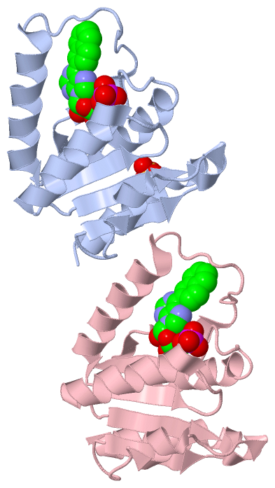
(-) Ligands, Modified Residues, Ions  (2, 3)

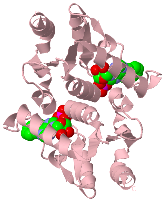
Asymmetric Unit (2, 3)
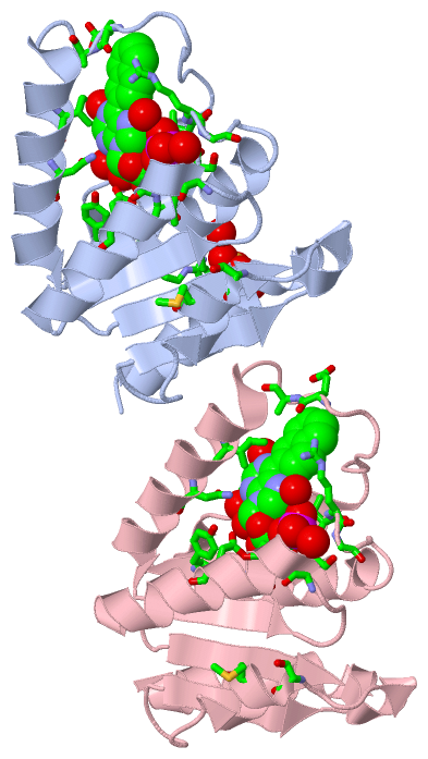
No.NameCountTypeFull Name
1N6P2Ligand/Ion6-(NAPHTHALEN-2-YL)-9-(5-O-PHOSPHONO-BETA-D-RIBOFURANOSYL)-9H-PURINE
2SO41Ligand/IonSULFATE ION
Biological Unit 1 (1, 2)
No.NameCountTypeFull Name
1N6P2Ligand/Ion6-(NAPHTHALEN-2-YL)-9-(5-O-PHOSPHONO-BETA-D-RIBOFURANOSYL)-9H-PURINE
2SO4-1Ligand/IonSULFATE ION
Biological Unit 2 (2, 4)
No.NameCountTypeFull Name
1N6P2Ligand/Ion6-(NAPHTHALEN-2-YL)-9-(5-O-PHOSPHONO-BETA-D-RIBOFURANOSYL)-9H-PURINE
2SO42Ligand/IonSULFATE ION

(-) Sites  (3, 3)

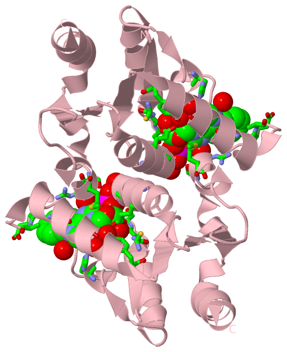
Asymmetric Unit (3, 3)
No.NameEvidenceResiduesDescription
1AC1SOFTWARETYR C:13 , CYS C:15 , GLY C:16 , SER C:17 , ILE C:18 , ARG C:19 , GLY C:20 , HIS C:45 , GLU C:57 , ALA C:58 , ILE C:65 , ASN C:69 , SER C:87 , GLY C:89 , GLU C:93 , SER C:117 , MET C:119 , HOH C:304 , HOH C:306 , HOH C:330 , HOH C:344binding site for residue N6P C 201
2AC2SOFTWARETYR A:13 , CYS A:15 , GLY A:16 , SER A:17 , ILE A:18 , ARG A:19 , GLY A:20 , HIS A:45 , VAL A:46 , GLU A:57 , ALA A:58 , ILE A:65 , ASN A:69 , SER A:87 , GLY A:89 , VAL A:90 , GLU A:93 , SER A:117 , MET A:119 , HOH A:305 , HOH A:315 , HOH A:369 , HOH A:385binding site for residue N6P A 201
3AC3SOFTWAREARG A:34 , ARG A:36 , ARG A:37 , HOH A:304 , HOH A:317 , HOH A:382binding site for residue SO4 A 202

(-) SS Bonds  (0, 0)

(no "SS Bond" information available for 4P5D)

(-) Cis Peptide Bonds  (0, 0)

(no "Cis Peptide Bond" information available for 4P5D)

 Sequence-Structure Mapping

(-) SAPs(SNPs)/Variants  (0, 0)

(no "SAP(SNP)/Variant" information available for 4P5D)

(-) PROSITE Motifs  (0, 0)

(no "PROSITE Motif" information available for 4P5D)

(-) Exons   (0, 0)

(no "Exon" information available for 4P5D)

(-) Sequences/Alignments

Asymmetric Unit
   Reformat: Number of residues per line =  ('0' or empty: single-line sequence representation)
  Number of residues per labelling interval =   
  UniProt sequence: complete  aligned part    
   Show mapping: SCOP domains CATH domains Pfam domains Secondary structure (by author)
SAPs(SNPs) PROSITE motifs Exons
(details for a mapped element are shown in a popup box when the mouse pointer rests over it)
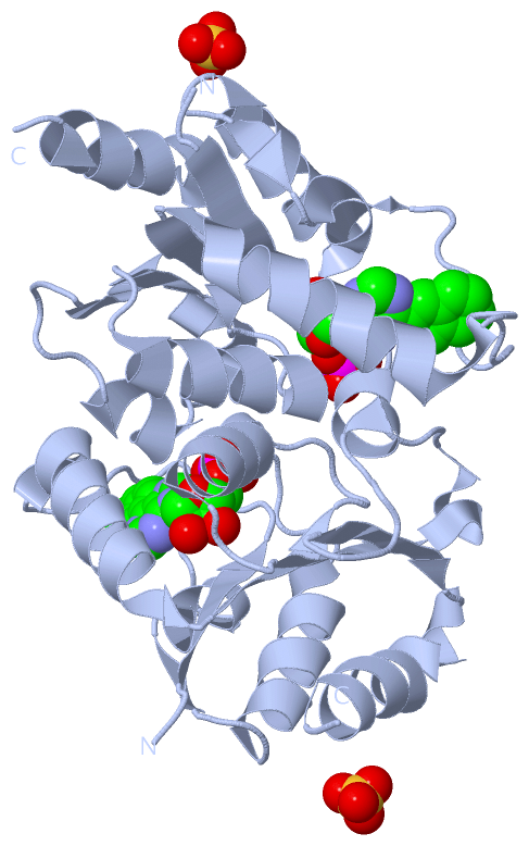
Chain A from PDB  Type:PROTEIN  Length:144
                                                                                                                                                                                
               SCOP domains ------------------------------------------------------------------------------------------------------------------------------------------------ SCOP domains
               CATH domains ------------------------------------------------------------------------------------------------------------------------------------------------ CATH domains
               Pfam domains ------------------------------------------------------------------------------------------------------------------------------------------------ Pfam domains
         Sec.struct. author ..eeeee........hhhhhhhhhhhhh...ee.hhhhhh...........hhhhhhhhhhhhhhhhh.eeeee....hhhhhhhhhhhhhh...eeeeehhhhh...hhhhhh......eeeee.hhhhhhhhhhhhhhhh.. Sec.struct. author
                 SAPs(SNPs) ------------------------------------------------------------------------------------------------------------------------------------------------ SAPs(SNPs)
                    PROSITE ------------------------------------------------------------------------------------------------------------------------------------------------ PROSITE
                 Transcript ------------------------------------------------------------------------------------------------------------------------------------------------ Transcript
                 4p5d A   9 RRSVYFCGSIRGGREDQALYARIVSRLRRYGKVLTEHVADAELEPLGEEAAGGDQFIHEQNLNWLQQADVVVAEVTQPSLGVGYELGRAVALGKPILCLFRPQSGRVLSAMIRGAADGSRFQVWDYAEGEVETMLDRYFEAYLV 152
                                    18        28        38        48        58        68        78        88        98       108       118       128       138       148    

Chain C from PDB  Type:PROTEIN  Length:142
                                                                                                                                                                              
               SCOP domains ---------------------------------------------------------------------------------------------------------------------------------------------- SCOP domains
               CATH domains ---------------------------------------------------------------------------------------------------------------------------------------------- CATH domains
               Pfam domains ---------------------------------------------------------------------------------------------------------------------------------------------- Pfam domains
         Sec.struct. author .eeeee........hhhhhhhhhhhhh...ee.hhhhhh...........hhhhhhhhhhhhhhhhh.eeeee....hhhhhhhhhhhhhhh..eeeeehhhhh...hhhhhh......eeeee.hhhhhhhhhhhhhhhh. Sec.struct. author
                 SAPs(SNPs) ---------------------------------------------------------------------------------------------------------------------------------------------- SAPs(SNPs)
                    PROSITE ---------------------------------------------------------------------------------------------------------------------------------------------- PROSITE
                 Transcript ---------------------------------------------------------------------------------------------------------------------------------------------- Transcript
                 4p5d C  10 RSVYFCGSIRGGREDQALYARIVSRLRRYGKVLTEHVADAELEPLGEEAAGGDQFIHEQNLNWLQQADVVVAEVTQPSLGVGYELGRAVALGKPILCLFRPQSGRVLSAMIRGAADGSRFQVWDYAEGEVETMLDRYFEAYL 151
                                    19        29        39        49        59        69        79        89        99       109       119       129       139       149  

   Legend:   → Mismatch (orange background)
  - → Gap (green background, '-', border residues have a numbering label)
    → Modified Residue (blue background, lower-case, 'x' indicates undefined single-letter code, labelled with number + name)
  x → Chemical Group (purple background, 'x', labelled with number + name, e.g. ACE or NH2)
  extra numbering lines below/above indicate numbering irregularities and modified residue names etc., number ends below/above '|'

 Classification and Annotation

(-) SCOP Domains  (0, 0)

(no "SCOP Domain" information available for 4P5D)

(-) CATH Domains  (0, 0)

(no "CATH Domain" information available for 4P5D)

(-) Pfam Domains  (0, 0)

(no "Pfam Domain" information available for 4P5D)

(-) Gene Ontology  (15, 15)

Asymmetric Unit(hide GO term definitions)

 Visualization

(-) Interactive Views

Asymmetric Unit
  Complete Structure
    Jena3D(integrated viewing of ligand, site, SAP, PROSITE, SCOP information)
    WebMol | AstexViewer[tm]@PDBe
(Java Applets, require no local installation except for Java; loading may be slow)
    STRAP
(Java WebStart application, automatic local installation, requires Java; full application with system access!)
    RasMol
(require local installation)
    Molscript (VRML)
(requires installation of a VRML viewer; select preferred view via VRML and generate a mono or stereo PDF format file)
 
  Ligands, Modified Residues, Ions
    N6P  [ RasMol | Jena3D ]  +environment [ RasMol | Jena3D ]
    SO4  [ RasMol | Jena3D ]  +environment [ RasMol | Jena3D ]
 
  Sites
    AC1  [ RasMol ]  +environment [ RasMol ]
    AC2  [ RasMol ]  +environment [ RasMol ]
    AC3  [ RasMol ]  +environment [ RasMol ]
 
  Cis Peptide Bonds
(no "Cis Peptide Bonds" information available for 4p5d)
 
Biological Units
  Complete Structure
    Biological Unit 1  [ Jena3D ]
    Biological Unit 2  [ Jena3D ]

(-) Still Images

Jmol
  protein: cartoon or spacefill or dots and stick; nucleic acid: cartoon and stick; ligands: spacefill; active site: stick
Molscript
  protein, nucleic acid: cartoon; ligands: spacefill; active site: ball and stick

 Databases and Analysis Tools

(-) Databases

Access by PDB/NDB ID
  4p5d
    Family and Domain InformationProDom | SYSTERS
    General Structural InformationGlycoscienceDB | MMDB | NDB | OCA | PDB | PDBe | PDBj | PDBsum | PDBWiki | PQS | PROTEOPEDIA
    Orientation in MembranesOPM
    Protein SurfaceSURFACE
    Secondary StructureDSSP (structure derived) | HSSP (homology derived)
    Structural GenomicsGeneCensus
    Structural NeighboursCE | VAST
    Structure ClassificationCATH | Dali | SCOP
    Validation and Original DataBMRB Data View | BMRB Restraints Grid | EDS | PROCHECK | RECOORD | WHAT_CHECK
 
Access by UniProt ID/Accession number
  DNPH1_RAT | O35820
    Comparative Protein Structure ModelsModBase
    Genomic InformationEnsembl
    Protein-protein InteractionDIP
    Sequence, Family and Domain InformationInterPro | Pfam | SMART | UniProtKB/SwissProt
 
Access by Enzyme Classificator   (EC Number)
  3.2.2.-
    General Enzyme InformationBRENDA | EC-PDB | Enzyme | IntEnz
    PathwayKEGG | MetaCyc
 
Access by Disease Identifier   (MIM ID)
  (no 'MIM ID' available)
    Disease InformationOMIM
 
Access by GenAge ID
  (no 'GenAge ID' available)
    Age Related InformationGenAge

(-) Analysis Tools

Access by PDB/NDB ID
    Domain InformationXDom
    Interatomic Contacts of Structural UnitsCSU
    Ligand-protein ContactsLPC
    Protein CavitiescastP
    Sequence and Secondary StructurePDBCartoon
    Structure AlignmentSTRAP(Java WebStart application, automatic local installation, requires Java; full application with system access!)
    Structure and Sequence BrowserSTING
 
Access by UniProt ID/Accession number
  DNPH1_RAT | O35820
    Protein Disorder PredictionDisEMBL | FoldIndex | GLOBPLOT (for more information see DisProt)

 Related Entries

(-) Entries Sharing at Least One Protein Chain (UniProt ID)

UniProtKB/Swiss-Prot
        DNPH1_RAT | O358202khz 2klh 4fyh 4fyi 4fyk 4kxl 4kxm 4kxn

(-) Related Entries Specified in the PDB File

4fyi