Show PDB file:   
         Plain Text   HTML   (compressed file size)
QuickSearch:   
by PDB,NDB,UniProt,PROSITE Code or Search Term(s)  
(-)Asymmetric Unit
(-)Asym. Unit - sites
(-)Biological Unit 1
(-)Biol. Unit 1 - sites
collapse expand < >
Image Asymmetric Unit
Asymmetric Unit  (Jmol Viewer)
Image Asym. Unit - sites
Asym. Unit - sites  (Jmol Viewer)
Image Biological Unit 1
Biological Unit 1  (Jmol Viewer)
Image Biol. Unit 1 - sites
Biol. Unit 1 - sites  (Jmol Viewer)

(-) Description

Title :  CRYSTAL STRUCTURE OF A/VICTORIA/361/2011 (H3N2) INFLUENZA VIRUS HEMAGGLUTININ
 
Authors :  P. S. Lee, I. A. Wilson
Date :  19 Dec 13  (Deposition) - 16 Apr 14  (Release) - 14 May 14  (Revision)
Method :  X-RAY DIFFRACTION
Resolution :  1.75
Chains :  Asym. Unit :  A,B
Biol. Unit 1:  A,B  (3x)
Keywords :  Viral Fusion Protein, Virus Attachment And Entry, Viral Protein (Keyword Search: [Gene Ontology, PubMed, Web (Google))
 
Reference :  P. S. Lee, N. Ohshima, R. L. Stanfield, W. Yu, Y. Iba, Y. Okuno, Y. Kurosawa, I. A. Wilson
Receptor Mimicry By Antibody F045-092 Facilitates Universal Binding To The H3 Subtype Of Influenza Virus.
Nat Commun V. 5 3614 2014
PubMed-ID: 24717798  |  Reference-DOI: 10.1038/NCOMMS4614

(-) Compounds

Molecule 1 - HEMAGGLUTININ HA1 CHAIN
    ChainsA
    EngineeredYES
    Expression SystemTRICHOPLUSIA NI
    Expression System Taxid7111
    GeneHA
    Organism ScientificINFLUENZA A VIRUS
    Organism Taxid1331560
    StrainA/SINGAPORE/H2011.447/2011(H3N2)
 
Molecule 2 - HEMAGGLUTININ HA2 CHAIN
    ChainsB
    EngineeredYES
    Expression SystemTRICHOPLUSIA NI
    Expression System Taxid7111
    GeneHA
    Organism ScientificINFLUENZA A VIRUS
    Organism Taxid1331560
    StrainA/SINGAPORE/H2011.447/2011(H3N2)

 Structural Features

(-) Chains, Units

  12
Asymmetric Unit AB
Biological Unit 1 (3x)AB

Summary Information (see also Sequences/Alignments below)

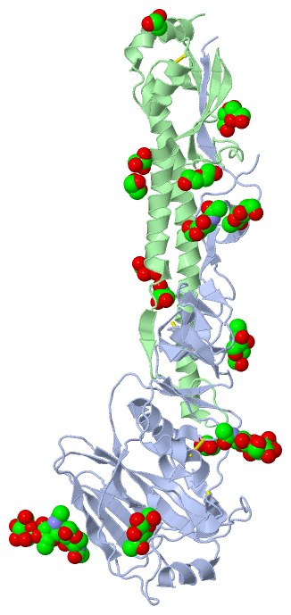
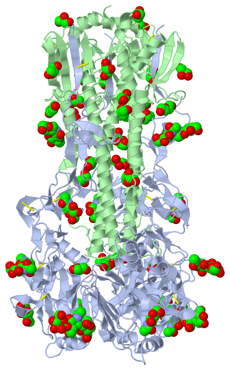
(-) Ligands, Modified Residues, Ions  (4, 18)

Asymmetric Unit (4, 18)
No.NameCountTypeFull Name
1BMA1Ligand/IonBETA-D-MANNOSE
2GOL8Ligand/IonGLYCEROL
3NAG8Ligand/IonN-ACETYL-D-GLUCOSAMINE
4PEG1Ligand/IonDI(HYDROXYETHYL)ETHER
Biological Unit 1 (4, 54)
No.NameCountTypeFull Name
1BMA3Ligand/IonBETA-D-MANNOSE
2GOL24Ligand/IonGLYCEROL
3NAG24Ligand/IonN-ACETYL-D-GLUCOSAMINE
4PEG3Ligand/IonDI(HYDROXYETHYL)ETHER

(-) Sites  (18, 18)

Asymmetric Unit (18, 18)
No.NameEvidenceResiduesDescription
01AC1SOFTWAREASN A:22 , ASN A:38 , HOH A:622 , HOH A:705BINDING SITE FOR RESIDUE NAG A 401
02AC2SOFTWAREASN A:38 , THR A:318 , LEU B:52BINDING SITE FOR RESIDUE NAG A 402
03AC3SOFTWAREGLU A:62 , ASN A:63 , TYR A:94 , NAG A:404 , HOH A:676BINDING SITE FOR RESIDUE NAG A 403
04AC4SOFTWARENAG A:403BINDING SITE FOR RESIDUE NAG A 404
05AC5SOFTWAREASN A:165 , SER A:219 , ARG A:222 , NAG A:406 , HOH A:717BINDING SITE FOR RESIDUE NAG A 405
06AC6SOFTWAREPRO A:221 , ARG A:222 , NAG A:405 , BMA A:407 , HOH A:717BINDING SITE FOR RESIDUE NAG A 406
07AC7SOFTWARENAG A:406BINDING SITE FOR RESIDUE BMA A 407
08AC8SOFTWAREASN A:45 , ASN A:285 , VAL A:297 , HOH A:665 , HOH A:668BINDING SITE FOR RESIDUE NAG A 408
09AC9SOFTWAREASN A:133BINDING SITE FOR RESIDUE NAG A 409
10BC1SOFTWAREPRO A:293 , ARG A:307 , HOH A:538 , HOH A:669 , GOL B:202 , HOH B:442BINDING SITE FOR RESIDUE GOL A 410
11BC2SOFTWARETYR A:105 , ARG A:208BINDING SITE FOR RESIDUE GOL A 411
12BC3SOFTWAREHIS A:18 , MET B:17 , VAL B:18 , GLY B:20 , TRP B:21 , HOH B:472 , HOH B:486BINDING SITE FOR RESIDUE PEG B 201
13BC4SOFTWAREGOL A:410 , ARG B:54 , LEU B:55 , HOH B:459BINDING SITE FOR RESIDUE GOL B 202
14BC5SOFTWAREVAL B:161 , TYR B:162 , GLU B:165 , HOH B:367BINDING SITE FOR RESIDUE GOL B 203
15BC6SOFTWAREARG B:32 , GLN B:34BINDING SITE FOR RESIDUE GOL B 204
16BC7SOFTWAREPHE B:9 , MET B:133 , GLY B:134 , ASN B:135 , HOH B:392BINDING SITE FOR RESIDUE GOL B 205
17BC8SOFTWAREALA B:43 , ASP B:46 , GLN B:47 , GOL B:207 , HOH B:395BINDING SITE FOR RESIDUE GOL B 206
18BC9SOFTWARELYS B:39 , ALA B:43 , GLU B:114 , LYS B:117 , GOL B:206 , HOH B:395BINDING SITE FOR RESIDUE GOL B 207

(-) SS Bonds  (6, 6)

Asymmetric Unit
No.Residues
1A:14 -B:137
2A:52 -A:277
3A:64 -A:76
4A:97 -A:139
5A:281 -A:305
6B:144 -B:148

(-) Cis Peptide Bonds  (1, 1)

Asymmetric Unit
No.Residues
1Ser A:54 -Pro A:55

 Sequence-Structure Mapping

(-) SAPs(SNPs)/Variants  (0, 0)

(no "SAP(SNP)/Variant" information available for 4O5N)

(-) PROSITE Motifs  (0, 0)

(no "PROSITE Motif" information available for 4O5N)

(-) Exons   (0, 0)

(no "Exon" information available for 4O5N)

(-) Sequences/Alignments

Asymmetric Unit
   Reformat: Number of residues per line =  ('0' or empty: single-line sequence representation)
  Number of residues per labelling interval =   
  UniProt sequence: complete  aligned part    
   Show mapping: SCOP domains CATH domains Pfam domains Secondary structure (by author)
SAPs(SNPs) PROSITE motifs Exons
(details for a mapped element are shown in a popup box when the mouse pointer rests over it)
Chain A from PDB  Type:PROTEIN  Length:317
                                                                                                                                                                                                                                                                                                                                                             
               SCOP domains ----------------------------------------------------------------------------------------------------------------------------------------------------------------------------------------------------------------------------------------------------------------------------------------------------------------------------- SCOP domains
               CATH domains ----------------------------------------------------------------------------------------------------------------------------------------------------------------------------------------------------------------------------------------------------------------------------------------------------------------------------- CATH domains
               Pfam domains ----------------------------------------------------------------------------------------------------------------------------------------------------------------------------------------------------------------------------------------------------------------------------------------------------------------------------- Pfam domains
         Sec.struct. author ..eeeeeee......eee.......eee..eee.ee......eeee...eee....hhhhhhhhhhhhhhhh.....eeee..........ee..hhhhhhhhhhhhh...eee.............eeeeee..eee....eee..........eeeeee......eeeeeeeee..hhhhhhhhhh.....eeee....eeee...............eeeeeeeee....eeeeee...eeee.eeee......eeee....eeeee..eee..eee.....ee.......eee.ee......eee........ Sec.struct. author
                 SAPs(SNPs) ----------------------------------------------------------------------------------------------------------------------------------------------------------------------------------------------------------------------------------------------------------------------------------------------------------------------------- SAPs(SNPs)
                    PROSITE ----------------------------------------------------------------------------------------------------------------------------------------------------------------------------------------------------------------------------------------------------------------------------------------------------------------------------- PROSITE
                 Transcript ----------------------------------------------------------------------------------------------------------------------------------------------------------------------------------------------------------------------------------------------------------------------------------------------------------------------------- Transcript
                 4o5n A   9 PGATLCLGHHAVPNGTIVKTITNDQIEVTNATELVQNSSIGEICDSPHQILDGENCTLIDALLGDPQCDGFQNKKWDLFVERSKAYSNCYPYDVPDYASLRSLVASSGTLEFNNESFNWTGVTQNGTSSACIRRSNNSFFSRLNWLTHLNFKYPALNVTMPNNEQFDKLYIWGVHHPGTDKDQIFLYAQSSGRITVSTKRSQQAVIPNIGSRPRIRNIPSRISIYWTIVKPGDILLINSTGNLIAPRGYFKIRSGKSSIMRSDAPIGKCNSECITPNGSIPNDKPFQNVNRITYGACPRYVKQSTLKLATGMRNVPE 325
                                    18        28        38        48        58        68        78        88        98       108       118       128       138       148       158       168       178       188       198       208       218       228       238       248       258       268       278       288       298       308       318       

Chain B from PDB  Type:PROTEIN  Length:173
                                                                                                                                                                                                             
               SCOP domains ----------------------------------------------------------------------------------------------------------------------------------------------------------------------------- SCOP domains
               CATH domains ----------------------------------------------------------------------------------------------------------------------------------------------------------------------------- CATH domains
               Pfam domains ----------------------------------------------------------------------------------------------------------------------------------------------------------------------------- Pfam domains
         Sec.struct. author .....................eeeeeee..eeeeeehhhhhhhhhhhhhhhhhhhh...eee............hhhhhhhhhhhhhhhhhhhhhhhhhhhhhhhhhhhhhhhhhhhhhhhhhhhhh..eee....eeee....hhhhhhhhhh...hhhhhhhhhhhhhh.. Sec.struct. author
                 SAPs(SNPs) ----------------------------------------------------------------------------------------------------------------------------------------------------------------------------- SAPs(SNPs)
                    PROSITE ----------------------------------------------------------------------------------------------------------------------------------------------------------------------------- PROSITE
                 Transcript ----------------------------------------------------------------------------------------------------------------------------------------------------------------------------- Transcript
                 4o5n B   1 GIFGAIAGFIENGWEGMVDGWYGFRHQNSEGRGQAADLKSTQAAIDQINGKLNRLIGKTNEKFHQIEKEFSEVEGRIQDLEKYVEDTKIDLWSYNAELLVALENQHTIDLTDSEMNKLFEKTKKQLRENAEDMGNGCFKIYHKCDNACIGSIRNGTYDHDVYRDEALNNRFQI 173
                                    10        20        30        40        50        60        70        80        90       100       110       120       130       140       150       160       170   

   Legend:   → Mismatch (orange background)
  - → Gap (green background, '-', border residues have a numbering label)
    → Modified Residue (blue background, lower-case, 'x' indicates undefined single-letter code, labelled with number + name)
  x → Chemical Group (purple background, 'x', labelled with number + name, e.g. ACE or NH2)
  extra numbering lines below/above indicate numbering irregularities and modified residue names etc., number ends below/above '|'

 Classification and Annotation

(-) SCOP Domains  (0, 0)

(no "SCOP Domain" information available for 4O5N)

(-) CATH Domains  (0, 0)

(no "CATH Domain" information available for 4O5N)

(-) Pfam Domains  (0, 0)

(no "Pfam Domain" information available for 4O5N)

(-) Gene Ontology  (17, 34)

Asymmetric Unit(hide GO term definitions)
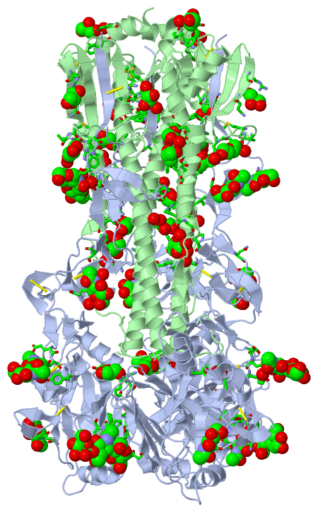
 Visualization

(-) Interactive Views

Asymmetric Unit
  Complete Structure
    Jena3D(integrated viewing of ligand, site, SAP, PROSITE, SCOP information)
    WebMol | AstexViewer[tm]@PDBe
(Java Applets, require no local installation except for Java; loading may be slow)
    STRAP
(Java WebStart application, automatic local installation, requires Java; full application with system access!)
    RasMol
(require local installation)
    Molscript (VRML)
(requires installation of a VRML viewer; select preferred view via VRML and generate a mono or stereo PDF format file)
 
  Ligands, Modified Residues, Ions
    BMA  [ RasMol | Jena3D ]  +environment [ RasMol | Jena3D ]
    GOL  [ RasMol | Jena3D ]  +environment [ RasMol | Jena3D ]
    NAG  [ RasMol | Jena3D ]  +environment [ RasMol | Jena3D ]
    PEG  [ RasMol | Jena3D ]  +environment [ RasMol | Jena3D ]
 
  Sites
    AC1  [ RasMol ]  +environment [ RasMol ]
    AC2  [ RasMol ]  +environment [ RasMol ]
    AC3  [ RasMol ]  +environment [ RasMol ]
    AC4  [ RasMol ]  +environment [ RasMol ]
    AC5  [ RasMol ]  +environment [ RasMol ]
    AC6  [ RasMol ]  +environment [ RasMol ]
    AC7  [ RasMol ]  +environment [ RasMol ]
    AC8  [ RasMol ]  +environment [ RasMol ]
    AC9  [ RasMol ]  +environment [ RasMol ]
    BC1  [ RasMol ]  +environment [ RasMol ]
    BC2  [ RasMol ]  +environment [ RasMol ]
    BC3  [ RasMol ]  +environment [ RasMol ]
    BC4  [ RasMol ]  +environment [ RasMol ]
    BC5  [ RasMol ]  +environment [ RasMol ]
    BC6  [ RasMol ]  +environment [ RasMol ]
    BC7  [ RasMol ]  +environment [ RasMol ]
    BC8  [ RasMol ]  +environment [ RasMol ]
    BC9  [ RasMol ]  +environment [ RasMol ]
 
  Cis Peptide Bonds
    Ser A:54 - Pro A:55   [ RasMol ]  
 
Biological Unit
  Complete Structure
    Biological Unit 1  [ Jena3D ]

(-) Still Images

Jmol
  protein: cartoon or spacefill or dots and stick; nucleic acid: cartoon and stick; ligands: spacefill; active site: stick
Molscript
  protein, nucleic acid: cartoon; ligands: spacefill; active site: ball and stick

 Databases and Analysis Tools

(-) Databases

Access by PDB/NDB ID
  4o5n
    Family and Domain InformationProDom | SYSTERS
    General Structural InformationGlycoscienceDB | MMDB | NDB | OCA | PDB | PDBe | PDBj | PDBsum | PDBWiki | PQS | PROTEOPEDIA
    Orientation in MembranesOPM
    Protein SurfaceSURFACE
    Secondary StructureDSSP (structure derived) | HSSP (homology derived)
    Structural GenomicsGeneCensus
    Structural NeighboursCE | VAST
    Structure ClassificationCATH | Dali | SCOP
    Validation and Original DataBMRB Data View | BMRB Restraints Grid | EDS | PROCHECK | RECOORD | WHAT_CHECK
 
Access by UniProt ID/Accession number
  A0A097PF39_9 | A0A097PF39
    Comparative Protein Structure ModelsModBase
    Genomic InformationEnsembl
    Protein-protein InteractionDIP
    Sequence, Family and Domain InformationInterPro | Pfam | SMART | UniProtKB/TrEMBL
  L0HR89_9INFA | L0HR89
    Comparative Protein Structure ModelsModBase
    Genomic InformationEnsembl
    Protein-protein InteractionDIP
    Sequence, Family and Domain InformationInterPro | Pfam | SMART | UniProtKB/TrEMBL
  R9U684_9INFA | R9U684
    Comparative Protein Structure ModelsModBase
    Genomic InformationEnsembl
    Protein-protein InteractionDIP
    Sequence, Family and Domain InformationInterPro | Pfam | SMART | UniProtKB/TrEMBL
 
Access by Enzyme Classificator   (EC Number)
  (no 'Enzyme Classificator' available)
    General Enzyme InformationBRENDA | EC-PDB | Enzyme | IntEnz
    PathwayKEGG | MetaCyc
 
Access by Disease Identifier   (MIM ID)
  (no 'MIM ID' available)
    Disease InformationOMIM
 
Access by GenAge ID
  (no 'GenAge ID' available)
    Age Related InformationGenAge

(-) Analysis Tools

Access by PDB/NDB ID
    Domain InformationXDom
    Interatomic Contacts of Structural UnitsCSU
    Ligand-protein ContactsLPC
    Protein CavitiescastP
    Sequence and Secondary StructurePDBCartoon
    Structure AlignmentSTRAP(Java WebStart application, automatic local installation, requires Java; full application with system access!)
    Structure and Sequence BrowserSTING
 
Access by UniProt ID/Accession number
  A0A097PF39_9 | A0A097PF39
    Protein Disorder PredictionDisEMBL | FoldIndex | GLOBPLOT (for more information see DisProt)
  L0HR89_9INFA | L0HR89
    Protein Disorder PredictionDisEMBL | FoldIndex | GLOBPLOT (for more information see DisProt)
  R9U684_9INFA | R9U684
    Protein Disorder PredictionDisEMBL | FoldIndex | GLOBPLOT (for more information see DisProt)

 Related Entries

(-) Entries Sharing at Least One Protein Chain (UniProt ID)

UniProtKB/TrEMBL
        A0A097PF39_9 | A0A097PF394o5i
        L0HR89_9INFA | L0HR894o5i
        R9U684_9INFA | R9U6844o5i 4we8 4we9 4wea

(-) Related Entries Specified in the PDB File

4o58 4o5i 4o5l