Show PDB file:   
         Plain Text   HTML   (compressed file size)
QuickSearch:   
by PDB,NDB,UniProt,PROSITE Code or Search Term(s)  
(-)Asym./Biol. Unit
(-)Asym./Biol. Unit - sites
collapse expand < >
Image Asym./Biol. Unit
Asym./Biol. Unit  (Jmol Viewer)
Image Asym./Biol. Unit - sites
Asym./Biol. Unit - sites  (Jmol Viewer)

(-) Description

Title :  TANDEM CHROMODOMAINS OF HUMAN CHD1 IN COMPLEX WITH INFLUENZA NS1 C-TERMINAL TAIL DIMETHYLATED AT K229
 
Authors :  S. Qin, C. Xu, W. Tempel, C. H. Arrowsmith, C. Bountra, A. M. Edwards, J. M Structural Genomics Consortium (Sgc)
Date :  18 Dec 13  (Deposition) - 16 Apr 14  (Release) - 04 Jun 14  (Revision)
Method :  X-RAY DIFFRACTION
Resolution :  1.87
Chains :  Asym./Biol. Unit :  A,B
Keywords :  Viral, Chromodomain, Structural Genomics, Structural Genomics Consortium, Sgc, Dna Binding Protein-Viral Protein Complex (Keyword Search: [Gene Ontology, PubMed, Web (Google))
 
Reference :  S. Qin, Y. Liu, W. Tempel, M. S. Eram, C. Bian, K. Liu, G. Senisterra, L. Crombet, M. Vedadi, J. Min
Structural Basis For Histone Mimicry And Hijacking Of Host Proteins By Influenza Virus Protein Ns1.
Nat Commun V. 5 3952 2014
PubMed-ID: 24853335  |  Reference-DOI: 10.1038/NCOMMS4952

(-) Compounds

Molecule 1 - CHROMODOMAIN-HELICASE-DNA-BINDING PROTEIN 1
    ChainsA
    EC Number3.6.4.12
    EngineeredYES
    Expression SystemESCHERICHIA COLI
    Expression System PlasmidPET28-MHL
    Expression System StrainBL21(DE3)-V2R-PRARE2
    Expression System Taxid469008
    FragmentUNP RESIDUES 268-443
    GeneCHD1
    Organism CommonHUMAN
    Organism ScientificHOMO SAPIENS
    Organism Taxid9606
    SynonymCHD-1, ATP-DEPENDENT HELICASE CHD1
 
Molecule 2 - NONSTRUCTURAL PROTEIN 1
    ChainsB
    EngineeredYES
    FragmentUNP RESIDUES 216-230
    Organism ScientificINFLUENZA A VIRUS
    Organism Taxid387159
    Other DetailsSYNTHETIC PEPTIDE
    SyntheticYES

 Structural Features

(-) Chains, Units

  12
Asymmetric/Biological Unit AB

Summary Information (see also Sequences/Alignments below)

(-) Ligands, Modified Residues, Ions  (3, 13)

Asymmetric/Biological Unit (3, 13)
No.NameCountTypeFull Name
1GOL1Ligand/IonGLYCEROL
2MLY1Mod. Amino AcidN-DIMETHYL-LYSINE
3UNX11Ligand/IonUNKNOWN ATOM OR ION

(-) Sites  (1, 1)

Asymmetric Unit (1, 1)
No.NameEvidenceResiduesDescription
1AC1SOFTWAREGLY A:324 , TRP A:325 , SER A:326 , HIS A:329 , SER A:422 , TRP A:423 , GLU A:424 , THR B:225 , ALA B:226 , ARG B:227BINDING SITE FOR RESIDUE GOL B 302

(-) SS Bonds  (0, 0)

(no "SS Bond" information available for 4O42)

(-) Cis Peptide Bonds  (0, 0)

(no "Cis Peptide Bond" information available for 4O42)

 Sequence-Structure Mapping

(-) SAPs(SNPs)/Variants  (0, 0)

(no "SAP(SNP)/Variant" information available for 4O42)

(-) PROSITE Motifs  (0, 0)

(no "PROSITE Motif" information available for 4O42)

(-) Exons   (0, 0)

(no "Exon" information available for 4O42)

(-) Sequences/Alignments

Asymmetric/Biological Unit
   Reformat: Number of residues per line =  ('0' or empty: single-line sequence representation)
  Number of residues per labelling interval =   
  UniProt sequence: complete  aligned part    
   Show mapping: SCOP domains CATH domains Pfam domains Secondary structure (by author)
SAPs(SNPs) PROSITE motifs Exons
(details for a mapped element are shown in a popup box when the mouse pointer rests over it)
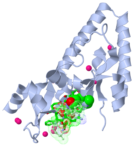
Chain A from PDB  Type:PROTEIN  Length:173
                                                                                                                                                                                                             
               SCOP domains ----------------------------------------------------------------------------------------------------------------------------------------------------------------------------- SCOP domains
               CATH domains ----------------------------------------------------------------------------------------------------------------------------------------------------------------------------- CATH domains
               Pfam domains ----------------------------------------------------------------------------------------------------------------------------------------------------------------------------- Pfam domains
         Sec.struct. author ....eeeeeeeeeee...hhhhhhhhhhhhhh............eeeeeeeee...hhhhheeehhhhhhhh....hhhhhhhhhhhhhhhhhhhh.hhhhhhhhhhhhhhhhhhhhhh..eeeeeee..........eeeeee...hhhhheeeehhhhhhhhhhhhhhhhh Sec.struct. author
                 SAPs(SNPs) ----------------------------------------------------------------------------------------------------------------------------------------------------------------------------- SAPs(SNPs)
                    PROSITE ----------------------------------------------------------------------------------------------------------------------------------------------------------------------------- PROSITE
                 Transcript ----------------------------------------------------------------------------------------------------------------------------------------------------------------------------- Transcript
                 4o42 A 270 EFETIERFMDCRIGRKGATGATTTIYAVEADGDPNAGFEKNKEPGEIQYLIKWKGWSHIHNTWETEETLKQQNVRGMKKLDNYKKKDQETKRWLKNASPEDVEYYNCQQELTDDLHKQYQIVERIIAHSNQKSAAGYPDYYCKWQGLPYSECSWEDGALISKKFQACIDEYFS 442
                                   279       289       299       309       319       329       339       349       359       369       379       389       399       409       419       429       439   

Chain B from PDB  Type:PROTEIN  Length:7
                                       
               SCOP domains ------- SCOP domains
               CATH domains ------- CATH domains
               Pfam domains ------- Pfam domains
         Sec.struct. author ....... Sec.struct. author
                 SAPs(SNPs) ------- SAPs(SNPs)
                    PROSITE ------- PROSITE
                 Transcript ------- Transcript
                 4o42 B 224 RTARSkV 230
                                 | 
                               229-MLY

   Legend:   → Mismatch (orange background)
  - → Gap (green background, '-', border residues have a numbering label)
    → Modified Residue (blue background, lower-case, 'x' indicates undefined single-letter code, labelled with number + name)
  x → Chemical Group (purple background, 'x', labelled with number + name, e.g. ACE or NH2)
  extra numbering lines below/above indicate numbering irregularities and modified residue names etc., number ends below/above '|'

 Classification and Annotation

(-) SCOP Domains  (0, 0)

(no "SCOP Domain" information available for 4O42)

(-) CATH Domains  (0, 0)

(no "CATH Domain" information available for 4O42)

(-) Pfam Domains  (0, 0)

(no "Pfam Domain" information available for 4O42)

(-) Gene Ontology  (27, 27)

Asymmetric/Biological Unit(hide GO term definitions)

 Visualization

(-) Interactive Views

Asymmetric/Biological Unit
  Complete Structure
    Jena3D(integrated viewing of ligand, site, SAP, PROSITE, SCOP information)
    WebMol | AstexViewer[tm]@PDBe
(Java Applets, require no local installation except for Java; loading may be slow)
    STRAP
(Java WebStart application, automatic local installation, requires Java; full application with system access!)
    RasMol
(require local installation)
    Molscript (VRML)
(requires installation of a VRML viewer; select preferred view via VRML and generate a mono or stereo PDF format file)
 
  Ligands, Modified Residues, Ions
    GOL  [ RasMol | Jena3D ]  +environment [ RasMol | Jena3D ]
    MLY  [ RasMol | Jena3D ]  +environment [ RasMol | Jena3D ]
    UNX  [ RasMol | Jena3D ]  +environment [ RasMol | Jena3D ]
 
  Sites
    AC1  [ RasMol ]  +environment [ RasMol ]
 
  Cis Peptide Bonds
(no "Cis Peptide Bonds" information available for 4o42)
 

(-) Still Images

Jmol
  protein: cartoon or spacefill or dots and stick; nucleic acid: cartoon and stick; ligands: spacefill; active site: stick
Molscript
  protein, nucleic acid: cartoon; ligands: spacefill; active site: ball and stick

 Databases and Analysis Tools

(-) Databases

Access by PDB/NDB ID
  4o42
    Family and Domain InformationProDom | SYSTERS
    General Structural InformationGlycoscienceDB | MMDB | NDB | OCA | PDB | PDBe | PDBj | PDBsum | PDBWiki | PQS | PROTEOPEDIA
    Orientation in MembranesOPM
    Protein SurfaceSURFACE
    Secondary StructureDSSP (structure derived) | HSSP (homology derived)
    Structural GenomicsGeneCensus
    Structural NeighboursCE | VAST
    Structure ClassificationCATH | Dali | SCOP
    Validation and Original DataBMRB Data View | BMRB Restraints Grid | EDS | PROCHECK | RECOORD | WHAT_CHECK
 
Access by UniProt ID/Accession number
  CHD1_HUMAN | O14646
    Comparative Protein Structure ModelsModBase
    Genomic InformationEnsembl
    Protein-protein InteractionDIP
    Sequence, Family and Domain InformationInterPro | Pfam | SMART | UniProtKB/SwissProt
  NS1_I83A8 | Q38SQ2
    Comparative Protein Structure ModelsModBase
    Genomic InformationEnsembl
    Protein-protein InteractionDIP
    Sequence, Family and Domain InformationInterPro | Pfam | SMART | UniProtKB/SwissProt
 
Access by Enzyme Classificator   (EC Number)
  3.6.4.12
    General Enzyme InformationBRENDA | EC-PDB | Enzyme | IntEnz
    PathwayKEGG | MetaCyc
 
Access by Disease Identifier   (MIM ID)
  (no 'MIM ID' available)
    Disease InformationOMIM
 
Access by GenAge ID
  (no 'GenAge ID' available)
    Age Related InformationGenAge

(-) Analysis Tools

Access by PDB/NDB ID
    Domain InformationXDom
    Interatomic Contacts of Structural UnitsCSU
    Ligand-protein ContactsLPC
    Protein CavitiescastP
    Sequence and Secondary StructurePDBCartoon
    Structure AlignmentSTRAP(Java WebStart application, automatic local installation, requires Java; full application with system access!)
    Structure and Sequence BrowserSTING
 
Access by UniProt ID/Accession number
  CHD1_HUMAN | O14646
    Protein Disorder PredictionDisEMBL | FoldIndex | GLOBPLOT (for more information see DisProt)
  NS1_I83A8 | Q38SQ2
    Protein Disorder PredictionDisEMBL | FoldIndex | GLOBPLOT (for more information see DisProt)

 Related Entries

(-) Entries Sharing at Least One Protein Chain (UniProt ID)

UniProtKB/Swiss-Prot
        CHD1_HUMAN | O146462b2t 2b2u 2b2v 2b2w 2b2y 2n39 4b4c 4nw2 5afw

(-) Related Entries Specified in the PDB File

(no "Related Entries Specified in the PDB File" available for 4O42)