|
|
|
|
 Description
|
|
Compounds
|
||||||||||||||||||||||||||||||||||||||||||||||||
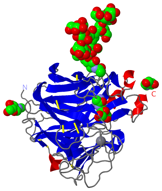
Chains, Units
Summary Information (see also Sequences/Alignments below) |
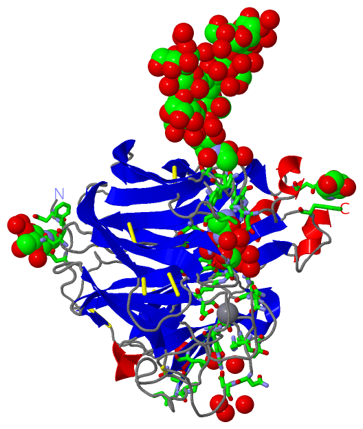
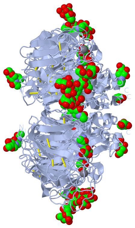
Ligands, Modified Residues, Ions (5, 13)| Asymmetric Unit (5, 13) Biological Unit 1 (4, 48) |
Sites (5, 5)
Asymmetric Unit (5, 5)
|
SS Bonds (9, 9)
Asymmetric Unit
|
||||||||||||||||||||||||||||||||||||||||
Cis Peptide Bonds (2, 2)
Asymmetric Unit
|
||||||||||||
SAPs(SNPs)/Variants (0, 0)| (no "SAP(SNP)/Variant" information available for 4MWR) |
PROSITE Motifs (0, 0)| (no "PROSITE Motif" information available for 4MWR) |
Exons (0, 0)| (no "Exon" information available for 4MWR) |
Sequences/Alignments
Asymmetric Unit
Chain A from PDB Type:PROTEIN Length:388
SCOP domains d4mwra_ A: automated matches SCOP domains
CATH domains ---------------------------------------------------------------------------------------------------------------------------------------------------------------------------------------------------------------------------------------------------------------------------------------------------------------------------------------------------------------------------------------------------- CATH domains
Pfam domains ---------------------------------------------------------------------------------------------------------------------------------------------------------------------------------------------------------------------------------------------------------------------------------------------------------------------------------------------------------------------------------------------------- Pfam domains
SAPs(SNPs) ---------------------------------------------------------------------------------------------------------------------------------------------------------------------------------------------------------------------------------------------------------------------------------------------------------------------------------------------------------------------------------------------------- SAPs(SNPs)
PROSITE ---------------------------------------------------------------------------------------------------------------------------------------------------------------------------------------------------------------------------------------------------------------------------------------------------------------------------------------------------------------------------------------------------- PROSITE
Transcript ---------------------------------------------------------------------------------------------------------------------------------------------------------------------------------------------------------------------------------------------------------------------------------------------------------------------------------------------------------------------------------------------------- Transcript
4mwr A 83 RNFNNLTKGLCTINSWHIYGKDNAVRIGESSDVLVTREPYVSCDPDECRFYALSQGTTIRGKHSNGTIHDRSQYRALISWPLSSPPTVYNSRVECIGWSSTSCHDGKSRMSICISGPNNNASAVVWYNRRPVAEINTWARNILRTQESECVCHNGVCPVVFTDGSATGPADTRIYYFKEGKILKWESLTGTAKHIEECSCYGERTGITCTCRDNWQGSNRPVIQIDPVAMTHTSQYICSPVLTDNPRPNDPNIGKCNDPYPGNNNNGVKGFSYLDGANTWLGRTISTASRSGYEMLKVPNALTDDRSKPIQGQTIVLNADWSGYSGSFMDYWAEGDCYRACFYVELIRGRPKEDKVWWTSNSIVSMCSSTEFLGQWNWPDGAKIEYFL 470
92 102 112 122 132 142 152 162 172 182 192 202 212 222 232 242 252 262 272 282 292 302 312 322 332 342 352 362 372 382 392 402 412 422 432 442 452 462
|
||||||||||||||||||||
SCOP Domains (1, 1)
Asymmetric Unit
 |
CATH Domains (0, 0)| (no "CATH Domain" information available for 4MWR) |
Pfam Domains (0, 0)| (no "Pfam Domain" information available for 4MWR) |
Gene Ontology (0, 0)|
Asymmetric Unit(hide GO term definitions)
(no "Gene Ontology" information available for 4MWR)
|
Interactive Views
|
||||||||||||||||||||||||||||||||||||||||||||||||||||||||||||||||||||||||||||||||||||||||||||||||||||||||||||||||||||||||||||||||||||||||||||||||||||||||||||||||||||||||||||||||||||||||||||||||||||||||
Still Images
|
||||||||||||||||
Databases
|
||||||||||||||||||||||||||||||||||||||||||||||||||||||||||||||||||||||||||||||||||||||||||||||||||||||||||||||||||||||||||||||||||||||||||||||||||||||||||||||||
Analysis Tools
|
|||||||||||||||||||||||||||||||||||||||||||||||||||||||||||||
Entries Sharing at Least One Protein Chain (UniProt ID)
Related Entries Specified in the PDB File
|
|